You must be signed in to read the rest of this article.
Registration on CDEWorld is free. You may also login to CDEWorld with your DentalAegis.com account.
This article reviews guidelines for construction of surgical guides to direct dental implant placement. Biologic principles are discussed with regard to the mesiodistal, buccolingual, and apicoronal insertion of implants. The application of these data to develop surgical guides is discussed. Also, with respect to specific prosthetic reconstructions, a variety of guides are described that can be fabricated for partially and fully edentulous arches.
Registration on CDEWorld is free. You may also login to CDEWorld with your DentalAegis.com account.
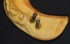
Figure 1
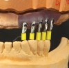
Figure 2
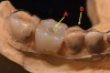
Figure 3
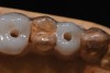
Figure 4
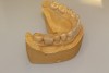
Figure 5
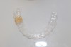
Figure 6
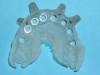
Figure 7
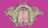
Figure 8
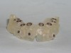
Figure 9
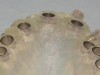
Figure 10
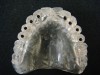
Figure 11
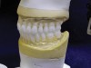
Figure 12
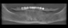
Figure 13
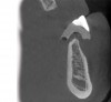
Figure 14
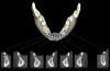
Figure 15
The author reports no conflicts of interest associated with this work.
Queries for the author may be directed to justin.romano@broadcastmed.com.