You must be signed in to read the rest of this article.
Registration on CDEWorld is free. You may also login to CDEWorld with your DentalAegis.com account.
Obtaining profound local anesthesia for patients receiving endodontic treatment can sometimes prove to be clinically challenging. By definition, local anesthesia causes a sensation loss in a specified area of the body produced by a depression of the excitation in nerve endings or an inhibition of the electrical action potential (impulses) that conducts along the nerve. Local anesthesia does not cause any loss of patient consciousness.1 Of the different theories regarding where local anesthesia works in the body, the specific receptor theory is most favored. This theory states that the local anesthesia binds to the specific receptors on the sodium channel.2 By blocking the sodium channel, the nerve’s electrical action potential is unable to fire its impulse.
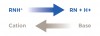
Because, in a clinical dental scenario, local anesthesia cannot be applied directly to a nerve membrane, its ability to diffuse through the tissue and cortical and cancellous bone is paramount. This is why clinicians need to allow more time for the local anesthesia to take effect. Also, the way in which the chemical dissociation of the local anesthesia occurs plays an important role in the ability of the anesthetic solution to penetrate the nerve tissue to block the nerve impulses. Local anesthestic solution is dispensed as salts (commonly hydrochloride salt) that are dissolved in either sterile water or saline. This solution has a base (RN) and a cation (RNH+) (Figure 1). The two main factors that enable the local anesthesia to be effective are diffusion of the anesthetic through the nerve sheath and the binding of the drug at the receptor site.3
The uncharged, lipid-soluble, free-base form (RN) diffuses through the nerve sheath. Once inside the sheath, a portion of the base (RN) reverts into the cation form (RNH+) through a re-equilibration process. This occurs because the base (RN) cannot exist in the RN form at a pH level of 7.4 (the normal pH level of the interior of a nerve cell). Approximately 75% of the base is converted to the cation (RNH+) inside the nerve sheath. It is the RNH+ cation that then is responsible for bonding to the sodium channel receptors and blocking the electrical action potential of the nerve conduction pathway.3
The amount of base (RN) or cation (RHN+) available through the dissociation of the local anesthesia will have a direct effect on the ability of the local anesthetic solution to clinically achieve profound anesthesia for the patient. The dissociation of local anesthesia and the relative proportions of each form (cation and base), therefore, are dependent on the pH level of the anesthetic solution and surrounding tissues. If a high pH level (fewer H+ ions) exists, the chemical reaction is driven to the right (Figure 1), thus providing more base than cation to penetrate the nerve sheath. If a low pH level (more H+ ions) exists, the chemical dissociation is driven to the left (Figure 1) and, therefore, more cation than base will be available, and, hence, the local anesthetic solution will have more impedance in penetrating the nerve sheath.4
Achieving Profound Anesthesia
Clinically, local anesthesia is achieved via the ability of the base (RN) to penetrate the nerve sheath. Inflammation, infection, and an anesthetics vasoconstrictor all play a role in lowering the pH measurement of the surrounding tissue. This low pH level decreases the amount of base available to penetrate the nerve sheath and, therefore, can delay or prevent local anesthetic from taking effect.5 In a study by Wallace et al,6 the authors speculated inflammation may alter the resting potentials and reduce the threshold of nerve excitability. This is commonly seen in patients receiving endodontic treatment who present with a pulpal diagnosis of symptomatic irreversible pulpitis.
The difficulties clinicians have in consistently achieving profound pulpal anesthesia on patients receiving endodontic treatment stem from a lack of understanding the clinical methodology, which needs to be followed prior to and after the administration of local anesthesia for endodontic treatment. The first common problem is that most clinicians do not objectively test teeth before and after the administration of local anesthesia to determine if pulpal anesthesia has been achieved. Instead, they make a clinical determination that pulpal anesthesia has been achieved based solely on the “subjective” viewpoint of the patient. If a patient states that his or her lips and/or cheeks are “numb” or “feel fat,” the clinician will often mistake these subjective signs and symptoms for having achieved pulpal anesthesia and will proceed with endodontic treatment. Unfortunately, by not objectively testing for pulpal anesthesia, a dentist may encounter an immediate pain response from the patient upon initiation of treatment. Although the patient’s subjective view helps in determining if the anesthesia is beginning to take effect, knowing whether a patient has reached a level of profound anesthetization needs to be based on “objective” testing performed by the clinician.
Objective Testing for Local Anesthesia Level
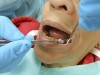
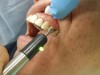
Prior to administering local anesthesia for endodontic treatment, the clinician needs to objectively test the treatment tooth with cold stimulus and/or an electric pulp tester (EPT) (Figure 2 and Figure 3). If the treatment tooth is nonresponsive to preoperative objective tests, the adjacent tooth should be tested. With a preoperative baseline reading of the pulp vitality, once anesthesia is “on board,” the level of anesthesia can be assessed by retesting the treatment tooth with cold stimulus or an EPT. If the postanesthesia tests result either in a negative response to cold or an EPT reading of 80, the patient will most likely be comfortable during the endodontic procedure.
Nusstein et al5 defined anesthetic success for mandibular anesthesia as achieving two consecutive EPT readings of 80 within 15 minutes and sustaining these readings for 60 minutes. In some cases of symptomatic irreversible pulpitis, the treatment teeth can objectively test negative to cold or have an EPT reading of 80 and yet the patient may still report pain on treatment.7,8 It is these case scenarios in which supplemental anesthesia injections need to be administered.
Supplemental Local Anesthesia
The use of soft-tissue anesthesia alone is not an effective predictor for pulpal anesthesia. Often clinically, a patient will report “lip numbness” after an inferior alveolar nerve block (IANB) or “cheek numbness” in a maxillary nerve block, and the tooth to be endodontically treated will either still test vital or not respond to vitality tests and yet still create patient discomfort on endodontic tooth access. Nusstein et al9 demonstrated that IANBs given to patients with irreversible pulpitis in a mandibular tooth have, on average, a 55% incidence of pulpal anesthesia, even in the presence of 100% lip numbness. To obtain pulpal anesthesia on these patients, the clinician must administer supplemental local anesthesia.
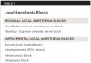
Prior to administering any supplemental anesthesia, the clinician must first administer local anesthesia for a “regional” block. For the mandibular region, a “regional” block is the IANB, and for maxillary teeth, it is the superior alveolar nerve block. Examples of supplemental local anesthesia injections are buccal nerve blocks (mandibular teeth), periodontal ligament (PDL), intraosseous, and intrapulpal (Table 1). If supplemental anesthesia is administered prior to a regional block, it will either be short acting or not effective enough to provide pulpal anesthesia.
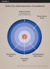
When administering supplemental nerve blocks, the clinician should start systematically with a buccal nerve block in mandibular teeth and a PDL block on maxillary teeth and progress the injection of anesthesia (as indicated) to an intrapulpal block. This “bull’s-eye” approach to the administration of anesthesia that progresses from regional to intrapulpal anesthesia is depicted in Figure 4.10
The supplemental anesthesia clinical methodology is as follows: After the anesthetic is injected into the buccal nerve for a mandibular tooth or the PDL of a maxillary tooth, the clinician should objectively test the treatment tooth with cold stimulus and/or EPT. If the mandibular or maxillary treatment tooth is nonresponsive to the test, the clinician should proceed with the endodontic treatment. If the patient, for any reason, still reports discomfort on access into the pulp chamber, an intrapulpal injection should be administered. When administering a mental nerve block (buccal block for the lower anterior teeth), empirically a clinician should have the patient apply hand pressure extraorally over the site of the mental nerve until the patient feels the further onset of the local anesthetic. This clinical technique will significantly reduce the onset time of the anesthesia.
If the mandibular tooth still tests vital in the above case scenario, a PDL injection should then be administered. This approach is based on a study by Matthews et al11 that demonstrated a buccal infiltration on patients with irreversible pulpitis reported a 58% success rate. If the maxillary treatment tooth still tests vital, a second PDL injection should be administered. A study by Cohen et al12 demonstrated that a reinjection of the PDL increased the success rate from 74% to 96% on teeth with a pretreatment pulpal diagnosis of irreversible pulpitis. Notably, the local anesthesia delivered from a PDL injection reaches the pulp by penetrating the cancellous bone through the natural osseous perforations in the tooth socket.13 The success of a PDL injection depends on the clinician’s ability to create backpressure during the injection.5
If a PDL injection in conjunction with a regional block and buccal nerve block in the case of a mandibular tooth does not achieve pulpal anesthesia, an intraosseous block should be considered. To a clinician who has never administered an intraosseous local anesthetic injection, it may seem like a challenging clinical task. However, with a didactic understanding and some clinical practice on easier cases, clinicians should find the intraosseous injection to be a valuable part of their local anesthesia technique. Several leading intraosseous injection systems are available on the market. Although each system varies slightly on specific clinical usage, the main similarities are that the injection should be administered distal to the tooth to be anesthetized, the anesthetic solution is placed directly into the cancellous bone surrounding the treatment tooth, and they all provide an immediate onset of local anesthesia after solution placement.5
Often, even after administering supplemental local anesthesia to a patient whose treatment tooth tests negative to cold stimulus and/or EPT, an intrapulpal injection will be necessary. Intrapulpal anesthesia is achieved as a result of pressure, not the actual type of anesthesia used.13 When performing an intrapulpal injection, a small cavity should be created in the pulp chamber and the anesthetic needle should fit “snugly” into this access. This will allow the anesthetic solution to be delivered under pressure. The patient should be advised that this procedure can be uncomfortable; however, the anesthesia will have a rapid onset and the patient will become comfortable with the initiation of endodontic treatment.14
In the event that a small-access cavity in the pulp chamber cannot be created due to clinical factors such as extensive decay, local anesthesia should be placed on the exposed pulp for about 1 minute before advancing the needle as far apically as possible into the pulp chamber so that the anesthetic solution is injected under pressure.14
Some endodontic treatment cases will present in which the clinician is able to access the pulp chamber and initiate file placement in the canal, but the patient will still report discomfort when the file advances in the apical third of the canal. Unfortunately, local anesthesia needles are too large to place anesthetic solution into this area. An alternative anesthesia technique is to place topical anesthetic on a No. 10 or No. 15 hand file and then advance the file into the apical third of the canal to effectively anesthetize and remove the vital pulp fragments. When using this technique, a slight amount of topical anesthetic should be applied to the hand file. Too much topical anesthetic can coat the canal walls and affect the sealing of the canal during obturation. Also, when the topical anesthetic mixes with sodium hypochlorite, it can create a darkish red and be mistaken for blood.15
Choosing the Correct Local Anesthetic
In conjunction with integrating regional and supplemental local anesthesia techniques, selecting the correct type of local anesthesia also plays a key role in obtaining profound pulpal anesthesia consistently and efficiently.
Regional Block
Dental schools have long taught that the more local anesthetic containing a vasoconstrictor (ie, epinephrine) that is administered clinically, the faster and more profound the local anesthetic effects will be for the patient. This methodology for administering an anesthetic with only a vasoconstrictor can actually delay the uptake of anesthetic in patients.10
Epinephrine is acidic and, therefore, lowers the pH level of the injection site. This lowered pH level (as stated earlier) will enable smaller amounts of base (RN) to be available for nerve-sheath penetration. Also, although epinephrine acts on both alpha receptors (vasoconstriction) and beta receptors (vasodilatation, bronchodilation, and increased heart rate and contraction), its effect on both is equal.16 This affects the amount of vasoconstriction that will actually occur in the injection site.
When giving a regional block—either an IANB or superior alveolar nerve block—the clinician should administer one carpule of an anesthetic with a vasoconstrictor and then wait a few minutes to observe if the patient reports any subjective signs (“feeling numb” or “feeling my lip or cheek is fat”). The first injection with an anesthetic with vasoconstrictor will help maintain the anesthetic in the region of the block.
If the patient states that he or she feels the anesthetic is taking effect, this indicates the clinician hit the anatomical block and can proceed with an anesthetic with no vasoconstrictor. If the patient does not report any subjective findings in regard to feeling numb, this means either the anatomical block was missed or an insufficient concentration of base was “on board” to penetrate the nerve sheath. Either way, the second carpule of anesthesia should still be 3% mepivacaine with no epinephrine. The subsequent injections with 3% mepivacaine will not further lower the surrounding pH level and, therefore, (as stated earlier) will enhance the amount of base (RN) to be available to penetrate the nerve sheath. After administering the second carpule of anesthetic, if the patient begins to feel numb, it will have been due to the anesthetic concentration; if the patient does not begin to feel numb, the anatomical placement of the anesthetic needs to be reevaluated.
Since the introduction of articaine into the US market, studies have demonstrated no significant difference between 4% articaine with 1:100,000 epinephrine and 2% lidociane with 1:100,000 epinephrine in IANB anesthesia.17,18 It is important to note that studies have reported a higher incidence of paresthesia when articaine has been administered for IANB. Although the exact etiology of the paresthesia is unknown, neurotoxicity may be due to the higher concentration of local anesthesia used: 4% articaine compared with 2% lidocaine.19,20 In a study by Yapp et al,21 they stated that the literature does not demonstrate a definitive relationship between the anesthetic agent and paresthesia. The clinician must consider these risks versus the benefits of administering 4% articaine for IANB anesthesia.
Buccal Block
When administering anesthesia for a buccal block, the use of 4% articaine with 1:100,000 epinephrine should be the drug of choice.18 A study by Srinivasan et al22 showed the efficacy of 4% articaine with 1:100,000 epinephrine was superior to 2% lidocaine with 1:100,000 epinephrine for buccal infiltrations in maxillary posterior teeth.
Periodontal Ligament Block
The use of 2% lidocaine with 1:100,000 epinephrine has been demonstrated to be significantly better in achieving pulpal anesthesia through a PDL injection as compared with using a local anesthetic without a vasoconstrictor.13,23
Intraosseous Block
The literature supports the use of an anesthetic with a vasoconstrictor for administering an intraosseous block;24 however, this will increase the heart rates in most patients.25 Therefore, clinicians need to balance a longer anesthetic effect accompanied by a tachycardia response of the patient versus using 3% mepivacaine (with no epinephrine) and eliminating the cardiac effect but shortening the duration of the local anesthetic effect. Reisman et al26 reported when a repeated intraosseous injection with 3% mepivacaine HCl (with no epinephrine) was administered, an increase in anesthetic success to 98% was achieved. Empirically, most patients feel uncomfortable when their hearts begin to race after the epinephrine systemically enters the system as the result of the intraosseous block.
Intrapulpal Block
As stated previously, the main objective in administering an intrapulpal anesthesia is to dispense it under pressure. It has been stated in the literature that saline is as effective as 2% lidocaine with 1:100,000 epinephrine in providing anesthesia intrapulpally.27,28 Empirically, the use of anesthetic with a vasoconstrictor will provide some vasoconstriction on the pulpal vascular system. This is important because most pulps with irreversible pulpitis can be hyperemic due to the body’s attempt to address the localized inflammation.
Summary
Clinicians need to recognize local anesthesia as a drug. Understanding how local anesthesia works, along with performing proper objective anesthesia level testing, administering supplemental anesthesia, and selecting the correct type of local anesthesia, will enable clinicians to enhance their consistency and efficiency in obtaining profound pulpal anesthesia on patients receiving endodontic treatment.
ABOUT THE AUTHORS
James Bahcall, DMD, MS
Clinical Associate Professor, Department of Endodontics,
University of Illinois-Chicago School of Dentistry, Chicago, Illinois; Diplomate, American Board of Endodontics
Qian Xie, DDS, PhD
Assistant Professor, Department of Endodontics, University of Illinois-Chicago School of Dentistry, Chicago, Illinois; Diplomate, American Board of Endodontics
Queries to the authors regarding this course may be submitted to authorqueries@aegiscomm.com.
REFERENCES
1. Malamed SF. Handbook of Local Anesthesia. 5th ed. St. Louis, MO: Elsevier Mosby; 2004:3.
2. Berry CA, Sanner JH, Keasling HH. A comparison of the anticonvulsant activity of mepivacaine and lidocaine. J Pharmacol Exp Ther. 1961;133(3):357-363.
3. Malamed SF. Handbook of Local Anesthesia. 5th ed. St. Louis, MO: Elsevier Mosby; 2004:16-24.
4. Malamed SF. Handbook of Local Anesthesia. 4th ed. St. Louis, MO: CV Mosby; 1997:14-17.
5. Nusstein JM, Reader A, Drum M. Local anesthesia strategies for the patient with a “hot” tooth. Dent Clin N Am. 2010;54(2):237-247.
6. Wallace JA, Michanowicz AE, Mundell RD, Wilson EG. A pilot study of the clinical problem of regionally anesthetizing the pulp of an acutely inflamed mandibular molar. Oral Surg Oral Med Oral Pathol. 1985;59(5):517-521.
7. Dreven LJ, Reader A, Beck M, et al. An evaluation of an electric pulp tester as a measure of analgesia in human vital teeth. J Endod. 1987;13 (5):233-238.
8. Hsiao-Wu GW, Susaria SM, White RR. Use of the cold test as a measure of pulpal anesthesia during endodontic therapy: a randomized, blinded, placebo-controlled clinical trial. J Endod. 2007;33(4):406-410.
9. Nusstein J, Reader A, Nist R, et al. Anesthetic efficacy of the supplemental intraosseous injection of 2% lidocaine with 1:100,000 epinephrine in irreversible pulpitis. J Endod. 1998;24(7):487-491.
10. Bahcall J. Everything I know about endodontics, I learned after dental school, Part 1. Dent Today. 2003;22(5):84-89.
11. Matthews R, Drum M, Reader A, et al. Articaine for supplemental buccal mandibular infiltration anesthesia in patients with irreversible pulpitis when the inferior alveolar nerve block fails. J Endod. 2009;35(3):343-346.
12. Cohen HP, Cha BY, Spångberg LS. Endodontic anesthesia in mandibular molars: a clinical study. J Endod. 1993;19(7):370-373.
13. Meechan JG. Supplementary routes to local anesthesia. Int Endod J. 2002;35(11):885-896.
14. Meechan JG. How to overcome failed local anesthesia. Br Dent J. 1999;186(1):15-20.
15. DeNunzio M. Topical anesthetic as an adjunct to local anesthesia during pulpectomies. J Endod. 1998;24(3):202-203.
16. Malamed SF. Handbook of Local Anesthesia. 5th ed. St. Louis, MO: Elsevier Mosby; 2004:45-49.
17. Brandt RG, Anderson PF, McDonald NJ, et al. The pulpal anesthetic efficacy of articaine versus lidocaine in dentistry: a meta-analysis. J Am Dent Assoc. 2011;142(5):493-504.
18. Mikesell P, Nusstein J, Reader A, et al. A comparison of articaine and lidocaine for inferior alveolar nerve blocks. J Endod. 2005;31(4):265-270.
19. Haas DA, Lennon D. A 21 year retrospective study of reports of paresthesia following local anesthetic administration. J Can Dent Assoc. 1995;61(4):319-330.
20. Garisto GA, Gaffen AS, Lawrence HP, et al. Occurrence of paresthesia after dental local anesthetic administration in the United States. J Am Dent Assoc. 2010;141(7):836-844.
21. Yapp KE, Hopcraft MS, Parashos P. Articaine: a review of the literature. Br Dent J. 2011;210(7):323-329.
22. Srinivasan N, Kavitha M, Loganathan C, Padmini G. Comparison of anesthetic efficacy of 4% articaine and 2% lidocaine for maxillary buccal infiltration in patients with irreversible pulpitis. Oral Surg Oral Med Oral Pathol Oral Radiol Endod. 2009;107(1):133-136.
23. Gray RJ, Lomax AM, Rood JP. Periodontal ligament injection: with or without a vasoconstrictor? Br Dent J. 1987;162(7):263-265.
24. Replogle K, Reader A, Nist R, et al. Anesthetic efficacy of the intraosseous injection of 2% lidocaine (1:100,000 epinephrine) and 3% mepivacaine in mandibular first molars. Oral Surg Oral Med Oral Pathol Oral Radiol Endod. 1997;83(1):30-37.
25. Coggins R, Reader A, Nist R, et al. Anesthetic efficacy of the intraosseous injection in maxillary and mandibular teeth. Oral Surg Oral Med Oral Pathol Oral Radiol Endod. 1996;81(6):634-641.
26. Reisman D, Reader A, Nist R, et al. Anesthetic efficacy of the supplemental intraosseous injection of 3% mepivacaine in irreversible pulpitis. Oral Surg Oral Med Oral Pathol Oral Radiol Endod. 1997;84(6):676-682.
27. Malamed SF. The management of pain and anxiety. In: Cohen S, Burns RC, eds. Pathways of the Pulp. 7th ed. St. Louis, MO: Mosby; 1998:665-666.
28. VanGheluwe J, Walton R. Intrapulpal injection: factors related to effectiveness. Oral Surg Oral Med Oral Pathol Oral Radiol Endod. 1997;83(1):38-40.