You must be signed in to read the rest of this article.
Registration on CDEWorld is free. You may also login to CDEWorld with your DentalAegis.com account.
Clinical investigators generally agree that scaling and root planing (SRP) effectively reduces the subgingival load of bacterial biofilms and dental calculus.1-3 However, SRP has its limitations. Numerous studies have reported that no one type of instrumentation (manual, sonic or ultrasonic scalers, or lasers) or technique (surgical or nonsurgical) is totally effective at eliminating all subgingival biofilm or calculus.1-19
There are multiple reasons for the inability to completely eradicate subgingival biofilms and calculus, the most common problems being access for adequate instrumentation and pocket probing depths (PPD). There is a direct correlation between increasing PPD and increasing presence of residual biofilms and calculus.1,3,5 Studies have reported that in pockets of 6 mm or greater, 19% to 66% of the root surface area will exhibit residual accretions.1,6,7,14-17 In pocket depths of 4 mm to 6 mm the contaminated root surface area drops to 15% to 38%.1,6,7,14-17 Of surprising interest, however, is the finding in several studies that root surfaces in shallow pockets, 0 mm to 3 mm in depth, commonly exhibit residual deposits of biofilm and calculus following SRP—the reported range being 4% to 43%.1,6,7,14-17 Additionally, it has been noted that following SRP, residual biofilms and calculus deposits are frequently noted on proximal surfaces and in furcations of multirooted teeth.2,5,18,19 Most surprising to many clinicians, however, are the reports that one of the more common areas to find post-SRP residual biofilm and calculus is the cemento-enamel junction (CEJ), an area that in most cases is easily accessible.3,5,9,14,20-22
Relationship of Cementum and Enamel at the CEJ
For more than a century23 the relationship of root cementum and coronal enamel at the CEJ has been addressed in multiple studies that used a variety of instrumentation methods, such as light microscopy,23-29 scanning electron microscopy,28,30-33 electron probe analysis,34-36 and embryologic and morphogenic analysis,37,38 to examine both healthy and diseased tissue. Although of great interest from a basic dental anatomy viewpoint, relatively few of these studies have addressed specific clinical implications of the various CEJ relationships.28,29,31,32,39 Indeed, none of the studies has addressed the question of why the CEJ is a potential biofilm and calculus trap.
Choquet (1899)23 was likely the first to publish a study of the CEJ. Numerous studies over the past century have reported on the variability of the relationship between enamel and cementum. Four such relationships have been reported: 1) enamel overlapping cementum; 2) cementum overlapping enamel; 3) an end-to-end (butt) joint; and 4) a gap between enamel and cementum that exposes underlying dentin.24-30,40,41 The prevalence of these relationships has been reported to vary considerably (Table 1). Indeed, it appears that multiple such relationships occur on individual teeth and on differing teeth within the same mouth, ie, incisor, cuspid, premolar, molar.28,30,40 As noted by Schroeder & Scherle,30 this latter fact renders the data from light microscopic studies rather useless, in terms of estimating the prevalence of the different cementum-enamel relationships, as the histologic section can only present the evidence for that specific area of sectioning. This, in turn, may contribute to the great variation in prevalence reported in various studies (Table 1).
Numerous authors have conjectured that the CEJ has become an area of increasing clinical interest due to the prevalence of cervical and root surface caries and lesions associated with abfraction and cervical abrasion.29,42-45 Aging trends in the US population indicate an increase in the dentate elderly46 and, as a consequence, one would expect a proportionate increase of CEJ involvement in restorative and periodontal problems. In this regard, Miller et al45 reported that 40.1% of abfraction lesions exhibited dental biofilm, 41.7% dental calculus, and 20.4% of teeth with abfractions also had bone loss due to periodontitis.
The CEJ as a Biofilm and Calculus Trap
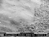
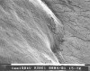
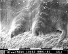
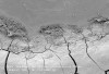
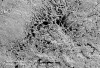
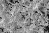
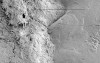
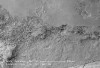
SEM examination of the surface textures of enamel (eg, hypoplastic enamel, perikymata), exposed dentin, and cementum at the CEJ and topographic contours of the various CEJ hard-tissue relationships reveals structural features that would appear to facilitate the attachment of bacterial biofilms and, if left undisturbed, the transition to dental calculus (Figure 1, Figure 2 , Figure 3 , Figure 4 , Figure 5 , Figure 6 , Figure 7 and Figure 8 ). Use of instrumentation in the CEJ area is difficult for several reasons. First, the irregular topography allows for biofilm development and calculus retention in depressions, which, in turn, requires significant root planing to remove all residual accretions. Second, the CEJ is often in close approximation to restoration margins that may inhibit adequate instrumentation due to overhangs or over-contouring at the gingival margins. Third, in the case of exposed dentin and dentinal tubules, instrumentation may cause patient discomfort, which, if local anesthesia is not administered, could cause the clinician to “let up” on the scaling and root planing. Lastly, as noted by several authors, specifically with reference to the deciduous dentition, the enamel at the CEJ is thin and relatively fragile.31,32 Although not reported in the literature, this same observation may be true of some adult teeth. Thus, in theory, aggressive instrumentation over time may result in fracturing of enamel and irregular contours at the CEJ ( Figure 9) that may contribute to biofilm and calculus retention.
Implications of Residual Biofilm at the CEJ
Biofilm attachment to the cervical enamel or root surface and subsequent maturation are key precursors to inflammatory periodontal disease. Subgingival biofilm, in different subjects, can exhibit considerable variation in amount and rate of accumulation and/or development. Such differences are influenced by host factors, such as xerostomia, nature of the surface presented for bacterial colonization, host genetics, etc; variations in oral hygiene; and bacterial composition.47,48
Biofilm adherence is highly influenced by surface texture and topography. Quirynen and van Steenberghe49 examined the relationship between early biofilm formation and surface topography and noted that on a smooth enamel surface the accumulating biofilm showed a preferential accumulation at the gingival margin with progressive development running parallel to the gingival margin. The authors further reported that when a groove was present on the tooth surface, biofilm growth was faster along the irregularity. The authors concluded that the pattern of biofilm growth was closely correlated to irregularities and roughness of the tooth surface.
The concept of surface roughness facilitating bacterial adhesion is related to surface-free energy.50-52 Simply put, increasingly rough surfaces exhibit increasing surface-free energy, and a high surface-free energy facilitates more bacterial adherence and, thereby, initiation of a biofilm. As described by Quirynen51 bacterial adhesion occurs in four phases: transport to the surface, initial adhesion with a reversible and irreversible stage, attachment by specific interactions, and colonization. Following colonization, the process of biofilm maturation, with incorporation of a diverse population of microbes, is a complex process involving multiple mechanisms, eg, binary fission, precipitation of new microbes onto the surface, intergeneric coaggregation, oxygen tensions within the mass, etc.53,54
Light and electron microscopic examinations of oral biofilms have demonstrated a high degree of order in colonization patterns.54 Cultural, immunologic or DNA probe assessments of subgingival biofilms have established that specific bacterial species frequently occur together in close association.54 In addition, cluster analysis of approximately 13,000 plaque samples has revealed that microbes exhibit a sequential appearance during colonization and a strong affinity to coaggregate.54 Socransky and Haffajee54,55 have described a sequence of six specific bacterial complexes—each assigned a color and each comprised of specific microbes—that interact during the process of successional colonization of both supra- and subgingival biofilms. The “orange” and “red” complexes, which appear later in the sequence of successional colonization, contain many of the better known periodontal pathogens, eg, Prevotella intermedia, Porphyromonas gingivalis, Tannerella forsythia, Treponema denticola, etc.55 Members of the red complex group (P gingivalis, T forsythia, T denticola) are rarely found in the absence of members from the orange complex group. With an increasing colonization by the orange complex, the red complex also increased in number, thus demonstrating bacterial interdependency, coaggregation, and succession colonization. The red complex species are associated with periodontal disease parameters such as deep periodontal probing depths and bleeding upon probing.54
As previously noted, numerous studies have confirmed that scaling and root planing can be an effective therapy for slight and moderate chronic periodontitis.3,56 However, it is also well established that not all biofilm and calculus is removed from subgingival root surfaces.5,7,17,20 In this regard, it is interesting to note that Caffesse et al5 reported that residual calculus was commonly found in deeper probing depths and most often in association with the CEJ.
Residual biofilms and calculus have important clinical ramifications. Several investigations have studied the relationship of supragingival biofilm to the re-colonization of subgingival biofilm following periodontal treatment. The microbes that re-colonize subgingival areas appear to have two possible origins: they may represent residual microbes following incomplete subgingival instrumentation, or they may be an extension of a growing and maturing supragingival biofilm. Repopulated subgingival biofilm is characterized by a dominant population of gram-negative anaerobes and motile bacteria—microbes generally associated with periodontal disease.57-59
Conclusion
With its variety of surface irregularities the CEJ presents a significant clinical challenge. The microscopic peaks and valleys, irregular enamel/cementum contours, possible presence of enamel hypoplasia, and the presence of subgingival perikymata provide unique niches that are sheltered from the shear forces inherent to oral hygiene and mastication. Further, CEJ contours may prevent adequate instrumentation, and yet over-instrumentation could potentially fracture the enamel and make the topography even more amenable to biofilm and calculus formation. Thus, the clinician is faced with a delicate balancing act regarding instrumentation of the CEJ region. Instrumentation must be as thorough as possible but not so aggressive as to create microfractures of the cervical enamel or ditching of cementum at the CEJ. Obviously, in this regard, using the tip of manual scalers or curettes or sonic and ultrasonic inserts should be considered with caution. The therapist must spend the time to be thorough but gentle as the CEJ may be fragile and susceptible to iatrogenic insult, which, in turn, can have long-term clinical implications regarding control and recurrence of periodontal disease.
References
1. Adriaens PA, Adriaens LM. Effects of nonsurgical periodontal therapy on hard and soft tissues. Periodontol 2000. 2004;36:121-145.
2. Claffey N, Polyzois I, Ziaka P. An overview of nonsurgical and surgical therapy. Periodontol 2000. 2004;36:35-44.
3. Cobb CM. Non-surgical pocket therapy: mechanical. Ann Periodontol. 1996;1(1):443-490.
4. Cobb CM, Low SB, Coluzzi DJ. Lasers and the treatment of chronic periodontitis. Dent Clin North Am. 2010;54(1):35-53. 5. Caffesse RG, Sweeney PL, Smith BA. Scaling and root planing with and without periodontal flap surgery. J Clin Periodontol. 1986;13(3):205-210.
6. Brayer WK, Mellonig JT, Dunlap RM, et al. Scaling and root planing effectiveness: the effect of root surface access and operator experience. J Periodontol. 1989;60(1)67-72.
7. Buchanan SA, Robertson PB. Calculus removal by scaling/root planing with and without surgical access. J Periodontol. 1987;58(3):159-163.
8. Dragoo MR. A clinical evaluation of hand and ultrasonic instruments on subgingival debridement. I. With unmodified and modified ultrasonic inserts. Int J Periodontics Restorative Dent. 1992;12(4):310-323.
9. Hunter RK, O’Leary TJ, Kafrawy AH. The effectiveness of hand versus ultrasonic instrumentation in open flap root planing. J Periodontol. 1984;55(12):697-703.
10. Nagy RJ, Otomo-Corgel J, Stambaugh R. The effectiveness of scaling and root planing with curets designed for deep pockets. J Periodontol. 1992;63(12):954-959.
11. Otero-Cagide FJ, Long BA. Comparative in vitro effectiveness of closed root debridement with fine instruments on specific areas of mandibular first molar furcations. II. Furcation area. J Periodontol. 1997;68(11):1098-1101.
12. Waerhaug J. Healing of the dento-epithelial junction following subgingival plaque control. II. As observed on extracted teeth. J Periodontol. 1978;49(3):119-134.
13. Wylam JM, Mealey BL, Mills MP, et al. The clinical effectiveness of open versus closed scaling and root planing on multi-rooted teeth. J Periodontol. 1993:64(11):1023-1028.
14. Gellin RG, Miller MC, Javed T, et al. The effectiveness of the Titan-S sonic scaler versus curettes in the removal of subgingival calculus. A human surgical evaluation. J Periodontol. 1986;57(11):672-680.
15. Rabbani GM, Ash MM Jr, Caffesse RG. The effectiveness of subgingival scaling and root planing in calculus removal. J Periodontol. 1981;52(3):119-123.
16. Rateitschak-Plüss EM, Schwarz JP, Guggenheim R, et al. Non-surgical periodontal treatment: where are the limits? An SEM study. J Clin Periodontol. 1992;19(4):240-244.
17. Sherman PR, Hutchens LH Jr, Jewson LG, et al. The effectiveness of subgingival scaling and root planing. I. Clinical detection of residual calculus. J Periodontol. 1990;61(1):3-8.
18. Matia JI, Bissada NF, Maybury JE, Ricchetti P. Efficiency of scaling of the molar furcation area with and without surgical access. Int J Periodontics Restorative Dent. 1986;6(6):24-35.
19. Stambaugh RV, Dragoo M, Smith DM, Carasali L. The limits of subgingival scaling. Int J Periodontics Restorative Dent. 1981;1(5):30-41.
20. Fleischer HC, Mellonig JT, Brayer WK, et al. Scaling and root planing efficacy in multirooted teeth. J Periodontol. 1989;60(7):402-409.
21. Breininger DR, O’Leary TJ, Blumenshine RV. Comparative effectiveness of ultrasonic and hand scaling for the removal of subgingival plaque and calculus. J Periodontol. 1987;58(1):9-18.
22. Patterson M, Eick JD, Eberhart AB, et al. The effectiveness of two sonic and two ultrasonic scaler tips in furcations. J Periodontol. 1989;60(6):325-329.
23. Choquet J. Note sur les rapports anatomiques existant chez l’homme entre l’émail et le cément. L’Odontologie. 1899;8:115-125.
24. Thorsen G. Tandems gingivale parti I forbindelse med nogen undersokeler over det anatomiske forhold mellem emalje og cement. Norske Tandlaegeform Tid. 1917;27:63-81.
25. Van Kirk LE. The frequency of the occurrence of certain structural variations in human enamel, dentin, and cementum. J Dent Res. 1928;8:459-461.
26. Birrer H. Zur Kenntnis der Schmelz-Zement-Zone des menschlichen Zahnes. Acta Anatomica. 1952;15:228-242.
27. Muller CJ, van Wyk CW. The amelo-cemental junction. J Dent Assoc S Afr. 1984;39(12):799-803.
28. Neuvald L, Consoiaro A. Cementoenamel junction: microscopic analysis and external cervical resorption. J Endod. 2000;26(9):503-508.
29. Arambawatta K, Peiris R, Nanayakkara D. Morphology of the cemento-enamel junction in premolar teeth. J Oral Sci. 2009;51(4):623-627.
30. Schroeder HE, Scherle WF. Cemento-enamel junction—revisted. J Periodont Res. 1988;23(1):53-59.
31. Francischone LA, Consolaro A. Morphology of the cementoenamel junction of primary teeth. J Dent Child. 2008;75(3):252-259.
32. Ceppi E, Dall’Oca S, Rimondini L, et al. Cementoenamel junction of deciduous teeth: SEM-morphology. Eur J Paediatr Dent. 2006; 7(3):131-134.
33. Akai M, Nakata T, Yamamoto K, et al. Scanning electron microscopy of cementoenamel junction. J Osaka Univ Dent Sch. 1978;18:83-94.
34. Atilla G, Baylas H. Electron probe analysis of cementum surfaces. J Marmara Univ Dent Fac. 1996;2(2-3):510-514.
35. Selvig KA, Hals E. Periodontally diseased cementum studied by correlated microradiography, electron probe analysis and electron microscopy. J Periodontal Res. 1977;12(6):419-429.
36. Barton NS, Van Swol RL. Periodontally diseased vs. normal roots as evaluated by scanning electron microscopy and electron probe analysis. J Periodontol. 1987;58(9):634-638.
37. Bosshardt DD, Schroeder HE. Cementogenesis reviewed: a comparison between human premolars and rodent molars. Anat Rec. 1996;245(2):267-292.
38. Owens PD. The root surface in human teeth: a microradiographic study. J Anat. 1976;122(pt 2):389-401.
39. Vacek JS, Gher ME. Cementum anomalies of the dentogingival junction. Int J Perio Rest Dent. 1993;13(5):443-449.
40. Grossman ES, Hargreaves JA. Variable cementoenamel junction in one person. J Prosthet Dent. 1991;65(1):93-97.
41. Avery JK. Histology of the periodontium: alveolar bone, cementum, and periodontal ligament. In: Avery JK, Steele PF, Avery N, ed. Oral Development and Histology, 3rd ed. New York, NY: Thieme; 2002:226-242.
42. Bevenius J, Lindskog S, Hultenby K. The amelocemental junction in young premolar teeth. A replica study by scanning electron microscopy. Acta Odontol Scand. 1993;51(3):135-142.
43. Beck JD, Hunt RJ, Hand JS, Field HM. Prevalence of root and coronal caries in a noninstitutionalized older population. J Am Dent Assoc. 1985;111(6):964-967.
44. Peltola P, Vehkalahti MM, Wuolijoki-Saaristo K. Oral health and treatment needs of the long-term hospitalised elderly. Gerodontology. 2004;21(2):93-99.
45. Miller N, Penaud, J, Ambrosini P, et al. Analysis of etiologic factors and periodontal conditions involved with 309 abfractions. J Clin Periodontol. 2003;30(9):828-832.
46. Centers for Disease Control and Prevention. MMWR Weekly. Public health and aging: retention of natural teeth among older adults - United States, 2002. 2003;52(50):1226-1229. Available at: www.cdc.gov/mmwr/preview/mmwrhtml. Accessed: June 21, 2010.
47. Socransky SS, Haffajee AD. Periodontal microbial ecology. Periodontol 2000. 2005;38:135-187.
48. Haffajee AD, Teles RP, Patel MR, et al. Factors affecting human supragingival biofilm composition. I. Plaque mass. J Periodontol Res. 2009;44(4):511-519.
49. Quirynen M, van Steenberghe D. Is early plaque growth rate constant with time? J Clin Periodontol. 1989;16(5):278-283.
50. Quirynen M, Marechal M, Busscher HJ, et al. The influence of surface free-energy on planimetric plaque growth in man. J Dent Res. 1989;68(5):796-799.
51. Quirynen M. The clinical meaning of the surface roughness and the surface free energy of intra-oral hard substrata on the microbiology of the supra- and subgingival plaque: results of in vitro and in vivo experiments. J Dent. 1994;22(suppl 1):S13-S16.
52. Quirynen M, Bollen CM. The influence of surface roughness and surface-free energy on supra and subgingival plaque formation in man. A review of the literature. J Clin Periodontol. 1995;22(1):1-14.
53. Cobb CM, Killoy WJ. Microbial colonization in human periodontal disease: an illustrated tutorial on selected ultrastructural and ecologic considerations. Scanning Microsc. 1990;4(3):675-691.
54. Socransky SS, Haffajee AD, Cugini MA, et al. Microbial complexes in subgingival plaque. J Clin Periodontol. 1998;25(2):134-144.
55. Haffajee AD, Socransky SS, Patel MR, Song X. Microbial complexes in supragingival plaque. Oral Microbiol Immunol. 2008;23(3):196-205.
56. Cobb CM. Clinical significance of non-surgical periodontal therapy: an evidence-based perspective of scaling and root planing. J Clin Periodontol. 2002;29(suppl 2):6-16.
57. Pedrazzoli V, Kilian M, Karring T, Kirkegaard E. Effect of surgical and non-surgical periodontal treatment on periodontal status and subgingival microbiota. J Clin Periodontol. 1991;18(8):598-604.
58. Mousques T, Listgarten MA, Phillips RW. Effect of scaling and root planing on the composition of the human subgingival microbial flora. J Periodont Res. 1980;15(2):144-151.
59. Sbordone L, Ramaglia L, Gulletta E, Iacono V. Recolonization of the subgingival microflora after scaling and root planing in human periodontitis. J Periodontol. 1990;61(9):579-584.
About the Authors
Keerthana Satheesh, BDS, DDS, MS
Clinical Associate Professor, Department of Periodontics School of Dentistry
University of Missouri-Kansas City
Kansas City, Missouri
Simon R. MacNeill, BDS, DDS
Associate Professor, Director of Graduate Periodontics Department of Periodontics
School of Dentistry
University of Missouri-Kansas City
Kansas City, Missouri
John W. Rapley, DDS, MS
Professor, Chair, Department of Periodontics
School of Dentistry
University of Missouri-Kansas City
Kansas City, Missouri
Charles M. Cobb, DDS, MS, PhD
Professor Emeritus, Department of Periodontics
School of Dentistry
University of Missouri-Kansas City
Kansas City, Missouri