You must be signed in to read the rest of this article.
Registration on CDEWorld is free. You may also login to CDEWorld with your DentalAegis.com account.
With approximately 5 million dental implants placed in the United States annually, these replacements for missing teeth are a well-established treatment modality with high long-term success rates.1 Peri-implant diseases, however, continue to pose a significant clinical challenge, as estimates of incidence rates for developing these conditions were about 53% for mucositis and 22% for peri-implantitis.2 Accurate diagnosis of peri-implant mucositis and peri-implantitis and a clear understanding of their multifactorial etiologies are essential for effective prevention and management of peri-implant diseases. Many patient-, site-, implant-, and restorative-related factors influence susceptibility to peri-implant complications, underscoring the importance of comprehensive risk assessment throughout implant treatment. This article reviews current diagnostic criteria and synthesizes the evidence on major risk indicators and risk factors to support informed clinical decision-making and promote long-term peri-implant health.
Diagnosis of Peri-implant Diseases and Conditions
Advances in dentistry, preventative care, and periodontal therapy have led to early disease detection resulting in decreased rates of tooth loss over time.3 While rates of complete edentulism are decreasing among adults, approximately 178 million patients in the United States are missing at least one tooth. Tooth replacement with dental implants is often optimal to achieve health, function, and esthetic outcomes.4 In 1999-2000, the percentage of individuals with dental implants in the United States was 0.7%; this increased to 5.7% in 2015-2016 and is expected to increase to up to 26% in 2026.3 As rates of dental implant placement rise, so have the rates of peri-implant disease. Accurate diagnosis and prevention of peri-implant diseases is necessary to ensure long-term dental implant success.5
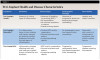
Peri-implant mucosa differs from gingival tissues around teeth in both blood supply and mechanisms of attachment.6 Dental implants do not have the connective tissue and periodontal ligament attachment seen around teeth; instead, they have a mucosal adherence, making them more susceptible to bacterial invasion and disease.7 Defining and characterizing peri-implant health and disease is critical to establish health and promote both primordial and primary prevention of peri-implant disease (Table 1).5,6,8
Patient-Related Implant Risk Factors
Systemic Conditions
Systemic conditions and autoimmune disorders such as diabetes mellitus, obesity, and rheumatoid arthritis are associated with increased risk of peri-implant disease.2,8 This may be due to altered healing potential and common elevated inflammatory burden. Systemic diseases can lead to increased oxidative stress, cytokine imbalance and cellular stress, which can cause elevated tissue destruction and impaired wound healing.2,8,9
Medications
Both proton pump inhibitors and selective serotonin reuptake inhibitors are associated with a 4.5% and 7.5% increased risk of early implant failure, respectively.10 Additionally, medications that result in altered host response, such as receptor activator of nuclear factor kappa-B (RANK) ligand inhibitors (denosumab), antiresorptive medications, nonsteroidal anti-inflammatory drugs, and corticosteroids, can impede implant bone healing and osseointegation.11
History of Periodontal Diseases
Active periodontitis (odds ratio [OR] ≈ 4.05) and/or a history of periodontal disease (OR ≈ 2.3) are risk factors for peri-implant disease.8,9,12 Similar destructive host response cascades to oral dysbiosis are seen in both periodontal and peri-implant disease.2,13,14 The common primary etiology and immune-inflammatory cellular activation result in parallel pathways for destruction at dental implants.2,9 Even in patients who have undergone successful periodontal treatment and have achieved periodontal stability, their demonstrated susceptibility makes it essential to adhere to a tight maintenance interval to control dysbiosis and modifiable risk factors.2,5,9,12
Nicotine Use
Nicotine use, including cigarette smoking, electronic nicotine delivery devices, and oral nicotine use, has been associated with increased risk of peri-implant disease, implant failure, and marginal bone loss (MBL).8,14 Nicotine use influences implant success through direct effects on vasoconstriction leading to decreased local oxygen and increased dysbiosis and indirect impacts on host immune-inflammatory response, cellular function, and tissue repair.15,16 Smokers demonstrate a two-fold risk for implant failure compared to nonsmokers, and each year of cessation reduces risk for periodontal and peri-implant attachment loss by 3.9%.5,6,17
Oral Hygiene and Peri-Implant Maintenance
Patient compliance and individualized care to facilitate regular, effective mechanical biofilm disruption is essential to achieving and maintaining peri-implant health. Because of the unique emergence profiles and contours of implant restorations, careful treatment planning for enhanced cleansability and use of a tailored oral home care armamentarium is critical for oral hygiene delivery. For example, electric toothbrushes demonstrate improved plaque control compared to manual toothbrushes at implant restorations.18,19 Further, interdental brushes, water flossers, end-tuft brushes, and specialized angled brushes may aid in plaque removal and should be individually tailored.20
Because dental implants are more susceptible to peri-implant disease compared to healthy teeth,9 risk-based peri-implant maintenance intervals should be utilized.21 An initial maintenance frequency of 3 to 4 months and periapical radiographs every 6 months for the first year after implant restoration is recommended to identify early signs of peri-implant disease and establish an initial implant-level diagnosis.8 Such close monitoring in the initial period after restoration allows dental healthcare professionals to establish baseline clinical parameters, detect peri-implant disease early, and provide initial intervention, if necessary.9
Site-Related Implant Risk Factors
Anatomical Considerations
Dental implants should be prosthetically driven and biologically executed. The use of digital technologies, including cone-beam computed tomography, intraoral scanning, and surgical guides, can improve surgical and prosthetic accuracy.22 Advanced imaging allows accurate identification of important anatomical structures, such as the maxillary sinuses and septa, mental foramina, inferior alveolar nerve, and lingual artery, and can decrease surgical morbidity during implant placement. It is critical, however, that ideal prosthetic rehabilitation and cleansability/maintenance be incorporated with anatomic considerations to ensure functional restorative care and long-term implant success. It has been reported that dental implants placed in the maxilla, particularly in the posterior, have higher failure rates than those placed in the mandible. This difference may be related to bone density and/or available bone volume.22,23
Bone Quality/Quantity
Sites with dense cortical bone (D1-D2), such as the anterior mandible, show increased initial implant stability and slower rates of bone loss compared to areas with higher trabecular bone (D3-D4), like the posterior maxilla.24
After tooth extraction without ridge preservation grafting, ridge resorption in both horizontal and vertical dimensions is likely to occur.25 Studies have demonstrated no significant difference in implant survival in grafted and/or native bone, and adequate bone thickness, particularly buccally, is predictive of both marginal bone and soft-tissue stability.26,27 Further, adequate bone volume can facilitate more ideal prosthetic contours and emergence angle, facilitating improved implant cleansability and long-term maintenance of peri-implant tissue stability.28
Soft-Tissue Quality/Quantity
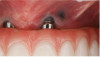
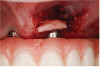
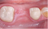
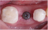
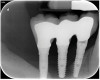
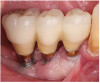
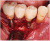
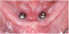
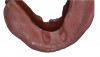
Soft-tissue quality and quantity play an important role in implant health and success. Implants with ≥2 mm keratinized tissue width and thick peri-implant phenotype demonstrate decreased inflammation, stable MBL, and improved esthetics.29,30 Soft-tissue augmentation, particularly free gingival graft or connective tissue graft (CTG) placement, may be recommended (Figure 1 through Figure 4). Such treatments can improve patient comfort during oral hygiene, reduce the risk of inflammation, and prevent future soft-tissue loss and recession around the implant.31
Previous Treatment and Infections
Active endodontic infections adjacent to and at planned implant sites have been associated with retrograde peri-implantitis and increased implant failure rates.25,32 It is suggested that these sites may continue to harbor pathogenic bacteria within the bone, putting the future implant at risk.32,33 Additionally, sites with a history of previous dental implant failure are associated with subsequent lower rates of cumulative implant success.34
Implant-Related Risk Factors
Implant Surface and Design
The most common dental implants placed today are made of titanium, titanium alloys, or zirconia, which have all been shown to be biocompatible. Differences in design and surface treatments significantly influence the biologic and mechanical environment surrounding the implant. Additionally, dental implant surfaces are often treated to create a roughened surface texture. Moderately roughened surfaces have demonstrated higher initial bone-to-implant contact compared to machined implant surfaces,35 but exposure of such roughened surfaces to the oral environment may result in increased plaque retention and progressive peri-implantitis.36
Implant Diameter and Length
Traditional thinking and focus on bone-to-implant contact led to placement of the widest, longest implants that were accommodated by the existing bone. Presently, it is understood that appropriate selection of implant size is critical for long-term success and that implant diameter and length may impact implant failure rates. Narrow-diameter dental implants (<3 mm) have been associated with lower levels of long-term survival compared to standard-diameter implants.37 However, when used appropriately and proper occlusion is managed, there is evidence to support the use of narrow-diameter implants.38 Both ultra-short (≤6 mm) and long (>13 mm) implants have been associated with increased failure rates.39,40 Further, when short implants (≥10 mm) are placed, wide-diameter implants have been associated with increased failure rates over the first 5 years.41 Identification of ideal implant size to appropriately replace missing teeth, accommodate occlusal forces, allow cleansability, and develop optimal emergence profiles is critical to long-term dental implant success.
Implant Positioning
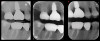
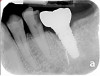
Improper 3-dimensional placement, including implants positioned too buccally, too apically, or too close to adjacent teeth or implants, can lead to thin or missing buccal bone, soft-tissue recession, and compromised restorations, leading to an increased risk of peri-implantitis (Figure 5 through Figure 9).9 Ideal implant placement includes placement with ≥1.5 mm to 2 mm of bone buccal and lingual to the proposed implant site, ≥3 mm between adjacent implant platforms, and ≥1.5 mm between an implant platform and adjacent teeth.9 Adequate inter-implant and tooth–implant spacing helps preserve crestal bone and supports stable papilla formation.5 Computer-assisted guided implant surgery has been shown to improve the ideal biologic and prosthetic implant placement compared to freehand placement.22 However, variables like pre-existing restorations, missing teeth, patient movement, poor moisture control, and inadequate tissue retraction can contribute to inaccuracies with intraoral scanning and 3-dimensional imaging.
Restorative and Occlusal Factors
Cement Vs Screw-Retained Restorations
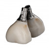
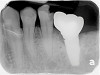
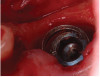
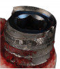
Excess cement around dental implants can trigger an inflammatory response leading to peri-implant disease42; however, evidence suggests that there is no significant difference in rates of peri-implantitis when comparing cement and screw-retained crowns if proper protocols for cementation are utilized.43,44 Decision-making for cement versus screw-retained restorations should take into account the margin position, as restorative margins located >1 mm subgingivally reduce the likelihood of complete cement removal.43 If margins are accessible, however, and adequate cement removal is achieved, resolution of peri-implant inflammation is seen in 75% of cases.44,45 Further, resin cements have shown increased rates of BOP, biofilm accumulation and difficulty in subgingival removal compared to zinc-oxide cements.46 Substitution of resin-based cement for zinc-oxide cement can lead to more than 75% reduction in BOP.46 Screw-retained restorations are sometimes favored for retrievability.42 Technical complications, such as screw loosening and porcelain fracture, are more common in screw-retained restorations compared to cement-retained, which can lead to peri-implant disease Figure 10 through Figure 13.44
Prosthetic Design
Many prosthetic design concerns are associated with malpositioned implant fixtures, highlighting the need for interdisciplinary treatment planning of the surgical and restorative approaches. An emergence angle of more than 30 degrees is associated with a two-fold increase in peri-implantitis.47 Such prostheses are associated with increased plaque accumulation and caries on adjacent teeth.48,49
Occlusal Issues
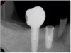
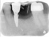
Oral parafunctional habits increase risk for prosthetic implant complications, including screw loosening, screw fracture, and implant and/or prosthetic fracture (Figure 14 through Figure 18).14 Once a prosthesis is loose, it can result in MBL and peri-implant disease. Implant failure rates are three to four times higher in bruxers compared to non-bruxers.21 It is suggested that heavy occlusal overload in conjunction with excessive eccentric forces results in microfractures in implant-supporting bone, disrupting bone resorption and apposition, leading to MBL.14 Careful design of protected occlusal schemes and verification of occlusal guard compliance is recommended for individuals with parafunctional habits, particularly given the low compliance rates that are documented for occlusal guard use.21
Clinical Decision Making
Clinical decision making for dental implant placement and restoration must begin with a comprehensive evaluation of the patient’s medical history, as systemic health directly influences healing capacity, local and systemic inflammatory burden, and long-term implant success. Systemic conditions such as dysglycemia, obesity, autoimmune disorders, and nicotine usage as well as select medications may influence initial healing and increase risk for peri-implant disease. Further, oral health, including periodontal disease, oral hygiene habits, and occlusal dysfunction, should also be identified and carefully considered during implant treatment planning. A detailed evaluation of patients’ current dental and medical conditions is necessary to ensure optimal implant treatment outcomes. Restorative plans should facilitate hygiene access and reduce biomechanical stress, especially in patients with parafunctional habits. Ultimately, implant therapy should only proceed when the patients’ medical conditions are stable, risks are clearly communicated, and the anticipated benefits outweigh potential complications (Figure 19).
Conclusion
Successful dental implant therapy relies on thoughtful clinical decision making that integrates the patient’s medical history with surgical and restorative planning. A thorough understanding of systemic conditions and their impact on healing, peri-implant disease risk, and long-term implant maintenance allows dental healthcare professionals to appropriately assess risk, modify treatment when necessary, and engage with interdisciplinary collaborators. By prioritizing medical stability, individualized risk assessment, and prosthetic designs that support hygiene and biomechanical balance, clinicians can improve predictability and optimize long-term implant outcomes.
ABOUT THE AUTHORS
Kallie Krueger, DMD
Periodontal Resident, University of Alabama at Birmingham School of Dentistry, Birmingham, Alabama
Maria L. Geisinger, DDS, MS
Professor and Chair, Kent and Phoebe Endowed Professor in Periodontology, Director, Advanced Education in Periodontology, Department of Periodontology, University of Alabama at Birmingham School of Dentistry, Birmingham, Alabama; Diplomate, American Board of Periodontology
Queries to the author regarding this course may be submitted to authorqueries@aegisdental.com.
REFERENCES
1. Boyce RA. Prosthodontic principles in dental implantology: adjustments in a coronavirus disease-19 pandemic-battered economy. Dent Clin North Am. 2021;65(1):135-165.
2. Galarraga-Vinueza ME, Pagni S, Finkelman M, et al. Prevalence, incidence, systemic, behavioral, and patient-related risk factors and indicators for peri-implant diseases: an AO/AAP systematic review and meta-analysis. J Periodontol. 2025;96(6):587-633.
3. Elani HW, Starr JR, Da Silva JD, Gallucci GO. Trends in dental implant use in the U.S., 1999-2016, and projections to 2026. J Dent Res. 2018;97(13):1424-1430.
4. Bidra A, Wu J. Talking points: missing teeth. American College of Prosthodontists. January 12, 2015. https://www.prosthodontics.org/assets/1/7/ACP_Talking_points_for_Missing_Teeth_1-12-15.pdf. Accessed October 2, 2026.
5. Heitz-Mayfield LJA, Salvi GE. Peri-implant mucositis. J Clin Periodontol. 2018;89 suppl 20:S257-S266.
6. Schwarz F, Derks J, Monje A, Wang HL. Peri-implantitis. J Periodontol. 2018;89 suppl 1:S267-S290.
7. Ivanovski S, Lee R. Comparison of peri-implant and periodontal marginal soft tissues in health and disease. Periodontol 2000. 2018;76(1):116-130.
8. Heitz-Mayfield LJA, Heitz F, Lang NP. Implant disease risk assessment IDRA–a tool for preventing peri-implant disease. Clin Oral Implants Res. 2020;31(4):397-403.
9. Wang HL, Avila-Ortiz G, Monje A, et al. AO/AAP consensus on prevention and management of peri-implant diseases and conditions: summary report. J Periodontol. 2025;96(6):519-541.
10. Chappuis V, Avila-Ortiz G, Araújo MG, Monje A. Medication-related dental implant failure: systematic review and meta-analysis. Clin Oral Implants Res. 2018;29 suppl 16:55-68.
11. Li JTW, Leung YY. Effect of antiresorptive drugs on osseointegrated dental implants: a systematic review. J Clin Med. 2024;13(7):2091.
12. Jepsen S, Berglundh T, Genco R, et al. Primary prevention of peri-implantitis: managing peri-implant mucositis. J Clin Periodontol. 2015;42 suppl 16:S152-S157.
13. Dahlen G, Lindhe J, Sato K, et al. The effect of supragingival plaque control on the subgingival microbiota in subjects with periodontal disease. J Clin Periodontol. 1992;19(10):802-809.
14. Chapple ILC, Van der Weijden F, Doerfer C, et al. Primary prevention of periodontitis: managing gingivitis. J Clin Periodontol. 2015;42 suppl 16:S71-S76.
15. Bredberg C, Vu C, Häggman-Henrikson B, Chrcanovic BR. Marginal bone loss around dental implants: comparison between matched groups of bruxer and non-bruxer patients: a retrospective case–control study. Clin Implant Dent Relat Res. 2023;25(1):124-132.
16. Chrcanovic BR, Albrektsson T, Wennerberg A. Smoking and dental implants: a systematic review and meta-analysis. J Dent. 2015;43(5):487-498.
17. Javed F, Rahman I, Romanos GE. Tobacco-product usage as a risk factor for dental implants. Periodontol 2000. 2019;81(1):48-56.
18. Renvert S, Persson GR, Pirih FQ, Camargo PM. Peri-implant health, peri-implant mucositis, and peri-implantitis: case definitions and diagnostic considerations. J Periodontol. 2018;89 suppl 1:S304-S312.
19. Van der Weijden GA, Timmerman MF, Nijboer A, et al. A comparative study of electric toothbrushes for the effectiveness of plaque removal in relation to toothbrushing duration. Timerstudy. J Clin Periodontol. 1993;20(7):476-481.
20. Van der Weijden FA, Slot DE. Efficacy of homecare regimens for mechanical plaque removal in managing gingivitis a meta review. J Clin Periodontol. 2015;42 suppl 16:S77-S91.
21. Sälzer S, Slot DE, Van der Weijden FA, Dörfer CE. Efficacy of inter-dental mechanical plaque control in managing gingivitis – a meta-review. J Clin Periodontol. 2015;42 suppl 16:S92-S105.
22. Curtis DA, Lin GH, Fishman A, et al. Patient-centered risk assessment in implant treatment planning. Int J Oral Maxillofac Implants. 2019;34(2):506-520.
23. Mahardawi B, Jiaranuchart S, Arunjaroensuk S, et al. The accuracy of dental implant placement with different methods of computer-assisted implant surgery: a network meta-analysis of clinical studies. Clin Oral Implants Res. 2025;36(1):1-16.
24. Lang LA, Hansen SE, Olvera N, Teich S. A comparison of implant complications and failures between the maxilla and the mandible. J Prosthet Dent. 2019;121(4):611-617.
25. Ortiz R, Maurício P, Mascarenhas PS. Densifying the future: a critical review of osseodensification and implant dentistry. Dent J (Basel). 2025;13(10):461.
26. Avila-Ortiz G, Chambrone L, Vignoletti F. Effect of alveolar ridge preservation interventions following tooth extraction: a systematic review and meta-analysis. J Clin Periodontol. 2019;46 suppl 21:195-223.
27. Farronato D, Mario Pasini P, Alain Orsina A, et al. Coorelation between buccal bone thickness at implant placement in healed sites and buccal soft tissue maturation pattern: a prospective three-year study. Materials (Basel). 2020;13(3):511.
28. Monje A, Roccuzzo A, Buser D, Wang H. Influence of buccal bone wall thickness on the peri-implant hard and soft tissue dimensional changes: a systematic review. Clin Oral Implants Res. 2023;34(3):157-176.
29. Izzetti R, Cinquini C, Nisi M, et al. Influence of prosthetic emergence profile on peri-implant marginal bone stability: a comprehensive review. Medicina (Kaunas). 2025;61(3):517.
30. Suárez-López Del Amo F, Lin GH, Monje A, et al. Influence of soft tissue thickness on peri-implant marginal bone loss: a systematic review and meta-analysis. J Periodontol. 2016;87(6):690-699.
31. Linkevicius T, Puisys A, Steigmann M, et al. Influence of vertical soft tissue thickness on crestal bone changes around implants with platform switching: a comparative clinical study. Clin Implant Dent Relat Res. 2015;17(6):1228-1236.
32. Zucchelli G, Tavelli L, Stefanini M, et al. Classification of facial peri-implant soft tissue dehiscence/deficiencies at single implant sites in the esthetic zone. J Periodontol. 2019;90(10):1116-1124.
33. Zhou W, Han C, Li D, et al. Endodontic treatment of teeth induces retrograde peri-implantitis. Clin Oral Implants Res. 2009;20(12):1326-1332.
34. Chrcanovic BR, Martins MD, Wennerberg A. Immediate placement of implants into infected sites: a systematic review. Clin Implant Dent Relat Res. 2015;17 suppl 1:e1-e16.
35. Oh SL, Shiau HJ, Reynolds MA. Survival of dental implants at sites after implant failure: a systematic review. J Prosthet Dent. 2020;123(1):54-60.
36. Romero-Serrano M, Romero-Ruiz MM, Herrero-Climent M, et al. Correlation between implant surface roughness and implant stability: a systematic review. Dent J (Basel). 2024;12(9):276.
37. Hussein A, Shah M, Atieh MA, et al. Influence of implant surfaces on peri-implant diseases – a systematic review and meta-analysis. Int Dent J. 2025;75(1):75-85.
38. Ortega-Oller I, Suárez F, Galindo-Moreno P, et al. The influence of implant diameter on its survival: a meta-analysis based on prospective clinical trials. J Periodontol. 2014;85(4):569-580.
39. Romanos GE, Schesni A, Nentwig GH, et al. Impact of implant diameter on success and survival of dental implants: an observational cohort study. Prosthesis. 2023;5(3):888-897.
40. Ravida A, Barootchi S, Askar H, et al. Long-term effectiveness of extra-short (≤ 6 mm) dental implants: a systematic review. Int J Oral Maxillofac Implants. 2019;34(1):68-84.
41. Jung RE, Al-Nawas B, Araujo M, et al. Group 1 ITI consensus report: the influence of implant length and design and medications on clinical and patient-reported outcomes. Clin Oral Implants Res. 2018;29 suppl 16:69-77.
42. Monje A, Fu JH, Chan HL, et al. Do implant length and width matter for short dental implants (<10 mm)? A meta-analysis of prospective studies. J Periodontol. 2013;84(12):1783-1791.
43. Sailer I, Mühlemann S, Zwahlen M, et al. Cemented and screw-retained implant reconstructions: a systematic review of the survival and complication rates. Clin Oral Implants Res. 2012;23 suppl 6:163-201.
44. Reis INRD, Fukuoka GL, Nagay BE, et al. Incidence of peri-implant disease associated with cement- and screw-retained implant-supported prostheses: a systematic review and meta-analysis. J Prosthet Dent. 2025;134(1):100-108.
45. Kotsakis GA, Zhang L, Gaillard P, et al. Investigation of the association between cement retention and prevalent peri-implant diseases: a cross-sectional study. J Periodontol. 2016;87(3):212-220.
46. Wilson TG Jr. The positive relationship between excess cement and peri-implant disease: a prospective clinical endoscopic study. J Periodontol. 2009;80(9):1388-1392.
47. Korsch M, Obst U, Walther W. Cement-associated peri-implantitis: a retrospective clinical observational study of fixed implant-supported restorations using a methacrylate cement. Clin Oral Implants Res. 2014;25(7):797-802.
48. Katafuchi M, Weinstein BF, Leroux BG, et al. Restoration contour is a risk indicator for peri-implantitis: a cross-sectional radiographic analysis. J Clin Periodontol. 2018;45(2):225-232.
49. Smith RB, Rawdin SB, Kagan V. Influence of implant-tooth proximity on incidence of caries in teeth adjacent to implants in molar sites: a retrospective radiographic analysis of 300 consecutive implants. Compend Contin Educ Dent. 2021;42(1):38-42.