You must be signed in to read the rest of this article.
Registration on CDEWorld is free. You may also login to CDEWorld with your DentalAegis.com account.
Autoimmune diseases are quite prevalent across the United States and can negatively affect oral health. A recent study using electronic health record data across six different medical systems reported that 4.6% of included patients were diagnosed with an autoimmune disease. Extrapolating for the US population of 333.3 million, this would estimate 15,440,225 individuals with an autoimmune disease.1 Traditional treatments involve immunosuppressant agents, including corticosteroids and methotrexate. Over the past several decades, biologic agents, a class of medications derived from living organisms designed to target specific aspects of the immune system and other cellular processes,2 have been developed for the management of autoimmune diseases.
Tumor necrosis factor-alpha (TNF-α) inhibitors are a notable subgroup of biologic agents used to manage certain autoimmune diseases characterized by chronic inflammation and tissue destruction. TNF-α is a cytokine that drives inflammation in various autoimmune conditions, including synovial inflammation and joint degradation in rheumatoid arthritis,3 intestinal mucosal damage in Crohn’s disease,4 and keratinocyte proliferation and skin lesion formation in psoriasis.5,6 TNF-α inhibitors bind TNF-α and prevent it from interacting with cell surface receptors. By targeting and neutralizing TNF-α, these inhibitors help alleviate the inflammatory responses that contribute to tissue damage and disease progression.7 These agents are widely prescribed, with several TNF-α inhibitors ranking among the most commonly prescribed drugs in the United States.8 While millions of patients use these agents, the impact on oral health and orthodontic treatment is still under investigation. TNF-α is known to regulate osteoclastogenesis and bone resorption, which are integral to orthodontic tooth movement (OTM).9 Therefore, inhibition of TNF-α could alter the biological response to orthodontic forces, potentially affecting treatment outcomes.
This article discusses the pharmacology of TNF-α inhibitors, their roles in bone metabolism and OTM, and the available evidence regarding the impact of TNF-α inhibitors on OTM. Understanding the intricate relationship between autoimmune diseases, their treatments, and oral health is essential for optimizing dental care. As orthodontists navigate the complexities associated with these conditions, awareness of potential complications and tailored treatment strategies becomes imperative to enhance patient outcomes and mitigate risks.
Pharmacology of TNF-α Inhibitors
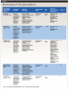
The clinical pharmacology of TNF-α inhibitors approved by the US Food and Drug Administration (FDA) is summarized in Table 1.10-14 These agents are all protein-based drugs, including several monoclonal antibodies,10-12 a fusion protein consisting of soluble TNF receptors,13 and a pegylated antibody fragment designed to enhance stability and reduce immunogenicity.14 As such, these drugs must be administered parenterally, either via intravenous infusion or subcutaneous injection, with dosing schedules tailored to the specific agent and the condition being treated. Numerous biosimilars have also been developed for these agents. Biosimilars are biologic agents developed to be highly similar to an approved reference product after the original drug patent has expired.15 Due to the inherent complexity of proteins, individual molecules may exhibit slight variations, making it impossible to create a chemically identical generic drug. Therefore, the FDA has established a dedicated regulatory pathway for biosimilars, which must demonstrate sufficient similarity in safety, efficacy, and potency to the reference product to enable their use as therapeutic alternatives.15
Despite their therapeutic benefits, TNF-α inhibitors are associated with a range of systemic adverse effects that warrant careful consideration.10-14 These effects are commonly categorized into local and immune-mediated reactions, infectious complications, and malignancy risks.
Injection site and infusion reactions are among the most frequently reported adverse events.16 These may include erythema, swelling, and discomfort at the administration site. Hypersensitivity reactions can also occur, ranging from delayed responses such as maculopapular exanthemas and urticaria to immediate immunoglobulin (Ig) E-mediated anaphylaxis.17 Immunogenicity varies among agents, with monoclonal antibodies generally posing a higher risk of anti-drug antibody formation than receptor fusion proteins.18
Another common adverse effect of TNF-α inhibitors is increased susceptibility to infections due to the immunosuppressant effects of the medications. Reactivation of latent tuberculosis infection, especially during the initial stages of treatment, is a well-established risk for patients undergoing TNF-α inhibitor therapy.19 As a result, screening for latent tuberculosis before initiating TNF-α inhibitors is recommended.12 Another concern with TNF-α inhibitor therapy is a relatively small yet notable risk for other serious opportunistic infections, including fungal and viral pathogens.20 Varicella zoster infection or reactivation in particular has been reported,21,22 so vaccination is recommended before starting therapy. Fungal infections may occur due to impaired mucosal immunity.23
Long-term use of TNF-α inhibitors has also been associated with a modest increased risk of certain malignancies, particularly skin cancers and non-Hodgkin lymphoma.24 However, autoimmune diseases themselves also predispose patients to various types of cancers, including lymphoma.25 While the absolute risk remains low, ongoing surveillance is advised, especially in patients with additional risk factors or prior malignancy history.26
The oral health effects associated with TNF-α inhibitor use are not well described. A recent scoping review explored the oral adverse effects associated with biologic agents in patients with inflammatory disorders.27 Of the included studies (N = 51) that reported on 17 biologic agents, oral adverse effects were most frequently reported for infliximab (17 publications), adalimumab (14 publications), and etanercept (9 publications). These adverse effects included angioedema, oral lichenoid lesions, medication-related osteonecrosis of the jaw, oral infections, oral squamous cell carcinoma, and other adverse effects such as orofacial granulomatosis, granulomatous ulceration, and oral ulcers. Notably, the majority of the included studies were case reports, so it is difficult to determine causation and the true prevalence of these oral adverse effects.
Due to the role of TNF-α in the development and progression of periodontal disease,28,29 it has been theorized that TNF-α inhibitors would improve periodontal disease status.30 Indeed, some studies have reported decreased gingival index, bleeding on probing, and probing depths31 and suggested improved healing with TNF-α inhibitor treatment.32 However, TNF-α inhibitor treatment has alternatively been reported to worsen gingival inflammation, potentially due to immune suppression.30 While there is no established association of TNF-α inhibitor treatment increasing the risk of orofacial infection, several cases of orofacial osteomyelitis following dental extraction have been reported.33,34 A single-center cross-sectional study reported that the decayed, missing, and filled tooth (DMFT) index was significantly higher in patients treated with biologic agents (59.7% treated with TNF-α inhibitors) compared to the average US adult index,31 suggesting that these agents may increase the risk of dental caries. Further research is necessary to confirm these associations and better understand the impact of TNF-α inhibitor treatment on oral health.
TNF-α in Orthodontic Tooth Movement
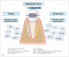
OTM involves a complex interplay between mechanical forces and biological responses. The application of orthodontic forces to the teeth induces cellular and inflammatory reactions in the surrounding periodontal tissues (Figure 1), ultimately leading to remodeling of the periodontal ligament (PDL) and alveolar bone, which facilitates tooth movement. Mechanical loading generates bone resorption on the compression side and new bone formation on the tension side. Bone resorption by osteoclasts, multinucleated cells derived from the monocyte/macrophage lineage, is the rate-limiting step in OTM. Osteoclasts adhere to the bone surface and degrade the mineralized matrix through enzymatic and acidic activity.
On the compression side, mechanical stress causes the release of proinflammatory mediators, including interleukin (IL)-1β, IL-6, IL-8, TNF-α, matrix metalloproteinase-1, prostaglandin E2 (PGE2), macrophage colony stimulating factor, and receptor activator of nuclear factor kappa-B ligand (RANKL).35,36 Conversely, on the tension side, tensile forces stimulate mesenchymal stem cells to differentiate into osteoblasts, which secrete bone matrix proteins and promote new bone formation.36 The newly formed matrix subsequently mineralizes, stabilizing the tooth in its new position.37 Osteogenic markers, including runt-related transcription factor 2, osteocalcin, and osteopontin, are upregulated, and higher levels of IL-10, tissue inhibitor of metalloproteinases-1, and collagen I are also observed on the tension side. Bone morphogenetic proteins (BMPs), particularly BMP-2 and BMP-4, further contribute by promoting osteoblast differentiation and enhancing bone formation under tensile loading.38
The role of TNF-α in OTM has been demonstrated in several studies. The levels of TNF-α in saliva increase during the early stages of orthodontic treatment and have been reported to be higher in patients treated with fixed appliances than in those treated with removable aligners.39 On the compression side, TNF-α plays a pivotal role in activating osteoclasts, the cells responsible for PDL widening and alveolar bone resorption.35,40-42 It directly amplifies RANKL expression in osteocytes.35,43 Together, TNF-α and RANKL synergistically promote osteoclast differentiation by increasing RANK+ precursor cells.44-48 Prostaglandins, particularly PGE2, further enhance this process by upregulating RANKL and downregulating osteoprotegerin, thereby promoting osteoclastogenesis. In addition, TNF-α upregulates osteocyte necroptosis on the compression side of OTM, leading to the release of damage-associated molecular patterns that further stimulate osteoclast activity.45 Evidence also indicates that TNF-α increases sclerostin levels, contributing to osteoclastic bone remodeling during OTM.41
Recent advances in periodontally accelerated osteogenic orthodontics, which promotes both osteoclastogenesis and osteogenesis to enhance tissue remodeling,36 have highlighted the regulatory role of TNF-α in modulating the rate of OTM. TNF-α facilitates this process by inducing transient acute physiological inflammation and promoting RANKL expression, thereby accelerating bone resorption on the compression side.49,50 Elevated TNF-α levels are also observed following localized bone injury techniques, such as corticotomy and micro-osteoperforation, which trigger the regional acceleratory phenomenon, a transient localized tissue response that enhances healing in both hard and soft tissues.51,52
Collectively, these findings underscore the critical role of TNF-α in mediating bone remodeling during OTM and suggest that targeted modulation of TNF-α signaling may influence treatment efficiency and outcomes.
Effect of TNF-α Inhibitors on Orthodontic Tooth Movement and Root Resorption
Although human data is lacking, several animal studies have investigated the effects of TNF-α inhibitors on OTM and root resorption. Jäger et al examined orthodontic bone remodeling in 80 male Wistar rats using a 0.5 N nickel–titanium (NiTi) coil spring for 12 days.53 Rats received daily intraperitoneal injections of soluble receptors to TNF-α, IL‑1, or both (2 ml of a 1 μg/ml solution) to inhibit the action of TNF-α and IL-1. Compared with controls, tooth movement decreased by approximately 50% in the cytokine‑receptor groups, with significantly fewer tartrate‑resistant acid phosphatase (TRAP)–positive osteoclasts and odontoclasts, less PDL hyalinization, and fewer resorption lacunae, indicating decreased bone and root resorption.
Similarly, systemic TNF-α blockade with infliximab suppresses OTM.54 Infliximab is a chimeric anti-TNF-α monoclonal antibody that binds both transmembrane and soluble TNF-α.55 In a rat model, weekly intraperitoneal infliximab (5 mg/kg for 4 weeks) significantly reduced OTM over 21 days (50 g NiTi coil spring), decreased osteoclast numbers on the compression side, lowered TNF-α, downregulated RANKL/RANK, and upregulated osteoprotegerin, indicating reduced osteoclastogenesis.
Hao and Hua evaluated dose, dosing interval, and delivery route of recombinant human soluble TNF-α receptor type I (rhsTNF‑RI) in a 14‑day rat OTM model (0.5 N, 72 male rats).56 Buccal submucoperiosteal rhsTNF-RI at 0.08–0.16 µg/mL significantly reduced tooth movement and TRAP-positive cells versus phosphate-buffered saline (PBS) control, whereas 0.04 µg/mL had no effect. Using 0.1 µg/mL, injections every 2 to 3 days were comparable to daily dosing, while every 4 days was inadequate; local (0.1 ml of 0.1 µg/ml every other day) and systemic (intraperitoneal injection, 1 ml of 1.0 µg/ml every other day) delivery produced similar inhibition. Overall, local injection of 0.1 mL at 0.1 µg/mL every 3 days was identified as an efficient regimen, supporting TNF-α blockade as a potential approach to prevent an orthodontic relapse and reduce resorptive activity, pending validation in human studies.
Zhang et al evaluated soluble TNF-α and IL-1 receptors in a rat model of mechanically induced root resorption.57 Using 18 male Wistar rats, localized trauma was created at the mesial gingiva/PDL of the maxillary first molar, and animals received daily intraperitoneal injections of soluble TNF-α receptor, soluble IL-1 receptor, or PBS (2 mL of 1 µg/mL), starting 1 day before injury. After 7 days, histologic and morphometric analyses showed root resorption was nearly abolished with TNF-α blockade, while IL-1 blockade produced only a moderate reduction, suggesting a more dominant role for TNF-α in odontoclast recruitment and activation in this model.
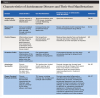
Taken together, these studies suggest that TNF-α inhibitors slow OTM and reduce root resorption, raising the possibility of their clinical application in preventing orthodontic relapse and severe root resorption. However, several factors limit clinical extrapolation. All experiments employed short‑term OTM models (<3 weeks), so the findings may not reflect responses in patients receiving long‑term TNF-α blockade during comprehensive treatment. The studies were conducted exclusively in male animals, leaving potential sex differences unaddressed, an important gap given the higher prevalence of autoimmune diseases in women.58 In addition, the animal models did not incorporate the autoimmune conditions for which TNF-α inhibitors are prescribed, making it difficult to generalize the results to affected patients, in whom the diseases themselves can influence oral health and orthodontic outcomes (Table 2).59-67 These limitations underscore the need for longer‑duration, sex‑stratified investigations, ideally including clinical cohorts, to evaluate the broader implications of both systemic for the treatment of autoimmune disease and locally delivered TNF-α inhibitors as an orthodontic therapeutic.
Conclusion
TNF-α plays a central role in OTM, promoting osteoclastogenesis and bone resorption on the compression side of the PDL. Modulating TNF-α signaling alters the rate of tooth movement, with implications for treatment efficiency, stability, and risks such as root resorption and relapse. Pharmacologically, TNF-α inhibitors are protein-based agents administered parenterally, with biosimilars increasingly becoming available. Despite their therapeutic benefits, these drugs carry risks of systemic adverse effects, including infections, hypersensitivity reactions, and malignancy, as well as oral complications such as angioedema, oral ulcers, and osteonecrosis of the jaw. Animal studies indicate that systemic or local TNF-α blockade can reduce OTM and root resorption by suppressing osteoclast activity and altering cytokine signaling. However, interpretation is constrained by short study durations, the absence of autoimmune disease models, and limited sex‑stratified analyses despite autoimmune conditions being more prevalent in women. Consequently, the clinical relevance for patients undergoing prolonged orthodontic treatment while receiving chronic TNF-α inhibitor therapy remains uncertain.
Understanding the complex interplay between autoimmune disease, biologic therapy, and orthodontic biomechanics is essential for dental professionals to optimize care, anticipate complications, and tailor treatment strategies for affected patients. Human studies, ideally incorporating disease phenotypes, medication exposure, duration, and sex‑specific analyses, are needed to define the long‑term impact of TNF‑α inhibitors on orthodontic outcomes and safety. Conducting retrospective case-controlled studies using existing clinical records, including patients undergoing orthodontic treatment with and without TNF-α inhibitor exposure, would be a crucial first step to evaluate potential differences in treatment duration, radiologic changes, and other relevant endpoints, as well as guide whether future prospective studies are warranted.
ABOUT THE AUTHORS
Neda Mahjour, DMD
Alumna, University of Pennsylvania School of Dental Medicine, Philadelphia, Pennsylvania
Elliot V. Hersh, DMD, MS, PhD
Professor, Department of Oral and Maxillofacial Surgery/Pharmacology, University of Pennsylvania School of Dental Medicine, Philadelphia, Pennsylvania
Hyeran Helen Jeon, DMD, MSD, DScD
Associate Professor of Orthodontics, Associate Director of Postdoctoral Orthodontics Program, Department of Orthodontics, University of Pennsylvania School of Dental Medicine, Philadelphia, Pennsylvania
Katherine France, DMD, MBE
Assistant Professor, Department of Oral Medicine, University of Pennsylvania School of Dental Medicine, Philadelphia, Pennsylvania
Katherine N. Theken, PharmD, PhD
Assistant Professor, Department of Oral and Maxillofacial Surgery/Pharmacology, University of Pennsylvania School of Dental Medicine, Philadelphia, Pennsylvania
Queries to the author regarding this course may be submitted to authorqueries@conexiant.com.
REFERENCES
1. Abend AH, He I, Bahroos N, et al. Estimation of prevalence of autoimmune diseases in the United States using electronic health record data. J Clin Invest. 2024;135(4):e178722.
2. US Food & Drug Administration. Frequently asked questions about therapeutic biological products. FDA website. Updated May 16, 2024. https://www.fda.gov/drugs/therapeutic-biologics-applications-bla/frequently-asked-questions-about-therapeutic-biological-products. Accessed September 3, 2026.
3. Komatsu N, Takayanagi H. Mechanisms of joint destruction in rheumatoid arthritis – immune cell-fibroblast-bone interactions. Nat Rev Rheumatol. 2022;18(7):415-429.
4. Noth R, Stüber E, Häsler R, et al. Anti-TNF-α antibodies improve intestinal barrier function in Crohn’s disease. J Crohns Colitis. 2012;6(4):464-469.
5. Mease P. TNFalpha therapy in psoriatic arthritis and psoriasis. Ann Rheum Dis. 2004;63(7):755-758.
6. Jang DI, Lee AH, Shin HY, et al. The role of tumor necrosis factor alpha (TNF-α) in autoimmune disease and current TNF-α inhibitors in therapeutics. Int J Mol Sci. 2021;22(5):2719.
7. Sedger LM, McDermott MF. TNF and TNF-receptors: from mediators of cell death and inflammation to therapeutic giants – past, present and future. Cytokine Growth Factor Rev. 2014;25(4):453-472.
8. Kane SP. ClinCalc DrugStats Database, Version 2025.08. https://clincalc.com/Drugstats. Accessed December 17, 2025.
9. Jeon HH, Teixeira H, Tsai A. Mechanistic insight into orthodontic tooth movement based on animal studies: a critical review. J Clin Med. 2021;10(8):1733.
10. AbbVie I. HUMIRA (adalimumab) injection, for subcutaneous use [package insert]. 2024.
11. Centocor Ortho Biotech I. SIMPONI (golimumab) prescribing information [package insert]. 2025.
12. Janssen Biotech I. REMICADE (infliximab) for injection, for intravenous use [package insert]. 2025.
13. Amgen I. ENBREL (etanercept) prescribing information [package insert]. 2024.
14. UCB Pharma I. CIMZIA (certolizumab pegol) prescribing information [package insert]. 2024.
15. US Food & Drug Administration. Biosimilars: review and approval. FDA website. Updated December 13, 2022. https://www.fda.gov/drugs/biosimilars/review-and-approval. Accessed September 3, 2026.
16. Gerriets V, Goyal A, Khaddour K. Tumor necrosis factor inhibitors. In: StatPearls [Internet]. Treasure Island, FL: StatPearls Publishing; 2024.
17. Puxeddu I, Caltran E, Rocchi V, et al. Hypersensitivity reactions during treatment with biological agents. Clin Exp Rheumatol. 2016;34(1):129-132.
18. Nesbitt A, Fossati G, Bergin M, et al. Mechanism of action of certolizumab pegol (CDP870): in vitro comparison with other anti-tumor necrosis factor alpha agents. Inflamm Bowel Dis. 2007;13(11):1323-1332.
19. Keane J, Gershon S, Wise RP, et al. Tuberculosis associated with infliximab, a tumor necrosis factor alpha-neutralizing agent. N Engl J Med. 2001;345(15):1098-1104.
20. Ali T, Kaitha S, Mahmood S, et al. Clinical use of anti-TNF therapy and increased risk of infections. Drug Healthc Patient Saf. 2013;5:79-99.
21. Kim BS, Maverakis E, Alexanian C, et al. Incidence, clinical features, management, and prevention of herpes zoster in patients receiving antitumor necrosis factor therapy: a clinical review. J Cutan Med Surg. 2020;24(3):278-284.
22. Strangfeld A, Listing J, Herzer P, et al. Risk of herpes zoster in patients with rheumatoid arthritis treated with anti-TNF-alpha agents. JAMA. 2009;301(7):737-744.
23. Filler SG, Yeaman MR, Sheppard DC. Tumor necrosis factor inhibition and invasive fungal infections. Clin Infect Dis. 2005;41 suppl 3:S208-S212.
24. D’Arcy ME, Beachler DC, Pfeiffer RM, et al. Tumor necrosis factor inhibitors and the risk of cancer among older Americans with rheumatoid arthritis. Cancer Epidemiol Biomarkers Prev. 2021;30(11):2059-2067.
25. Mercer LK, Galloway JB, Lunt M, et al. Risk of lymphoma in patients exposed to antitumour necrosis factor therapy: results from the British Society for Rheumatology Biologics Register for Rheumatoid Arthritis. Ann Rheum Dis. 2017;76(3):497-503.
26. Driscoll CB, Rich JM, Isaacson D, et al. Tumor necrosis factor-alpha inhibitor use and malignancy risk: a systematic review and patient level meta-analysis. Cancers (Basel). 2025;17(3):390.
27. France K, Yogarajah S, Gueiros LA, et al. World Workshop on Oral Medicine VII: oral adverse effects to biologic agents in patients with inflammatory disorders. A scoping review. J Oral Pathol Med. 2023;52(1):1-8.
28. Ding C, Ji X, Chen X, et al. TNF-α gene promoter polymorphisms contribute to periodontitis susceptibility: evidence from 46 studies. J Clin Periodontol. 2014;41(8):748-759.
29. Sumbayak IA, Masulili SLC, Tadjoedin FM, et al. Changes in interleukin-1β, tumor necrosis factor-α, and interleukin-10 cytokines in older people with periodontitis. Geriatrics (Basel). 2023;8(4):79.
30. Zhang J, Xu C, Gao L, et al. Influence of anti-rheumatic agents on the periodontal condition of patients with rheumatoid arthritis and periodontitis: a systematic review and meta-analysis. J Periodontal Res. 2021;56(6):1099-1115.
31. Shah S, Veerabagu N, Essel NAA, et al. The dental status of patients taking common biologic agents: a single-center cross-sectional study. Oral Dis. 2025;31(8):2641-2651.
32. Peddis N, Musu D, Ideo F, et al. Interaction of biologic therapy with apical periodontitis and periodontitis: a systematic review. Aust Dent J. 2019;64(2):122-134.
33. Ciantar M, Adlam DM. Treatment with infliximab: implications in oral surgery? A case report. Br J Oral Maxillofac Surg. 2007;45(6):507-510.
34. Bielory L. Immunobiologicals and ocular surface disease. Curr Opin Allergy Clin Immunol. 2022;22(5):314-318.
35. Marahleh A, Kitaura H, Ohori F, et al. Effect of TNF-α on osteocyte RANKL expression during orthodontic tooth movement. J Dent Sci. 2021;16(4):1191-1197.
36. Jeon HH, Huang X, Rojas Cortez L, et al. Inflammation and mechanical force-induced bone remodeling. Periodontol 2000. 2024;10.1111/prd.12619.
37. Garlet TP, Coelho U, Repeke CE, et al. Differential expression of osteoblast and osteoclast chemmoatractants in compression and tension sides during orthodontic movement. Cytokine. 2008;42(3):330-335.
38. Kamiya N, Mishina Y. New insights on the roles of BMP signaling in bone – a review of recent mouse genetic studies. Biofactors. 2011;37(2):75-82.
39. Karande P, BS R, Pandey A, et al. Study on evaluation of salivary tumor necrosis factor-alpha level in the treatment with fixed appliances and clear aligners. Int J Environmental Sci. 2025;11(15S):1461-1464.
40. Ogawa S, Kitaura H, Kishikawa A, et al. TNF-α is responsible for the contribution of stromal cells to osteoclast and odontoclast formation during orthodontic tooth movement. PLoS One. 2019;14(10):e0223989.
41. Ohori F, Kitaura H, Marahleh A, et al. Effect of TNF-α-induced sclerostin on osteocytes during orthodontic tooth movement. J Immunol Res. 2019;2019:9716758.
42. Alhashimi N, Frithiof L, Brudvik P, Bakhiet M. Orthodontic tooth movement and de novo synthesis of proinflammatory cytokines. Am J Orthod Dentofacial Orthop. 2001;119(3):307-312.
43. Marahleh A, Kitaura H, Ohori F, et al. TNF-α directly enhances osteocyte RANKL expression and promotes osteoclast formation. Front Immunol. 2019;10:2925.
44. Luo G, Li F, Li X, et al. TNF‑α and RANKL promote osteoclastogenesis by upregulating RANK via the NF‑κB pathway. Mol Med Rep. 2018;17(5):6605-6611.
45. Ohori F, Kitaura H, Marahleh A, et al. Osteocyte necroptosis drives osteoclastogenesis and alveolar bone resorption during orthodontic tooth movement. Sci Rep. 2025;15(1):19413.
46. Noguchi T, Kitaura H, Ogawa S, et al. TNF-α stimulates the expression of RANK during orthodontic tooth movement. Arch Oral Biol. 2020;117:104796.
47. Ubuzima P, Nshimiyimana E, Mukeshimana C, et al. Exploring biological mechanisms in orthodontic tooth movement: bridging the gap between basic research experiments and clinical applications – a comprehensive review. Ann Anat. 2024;255:152286.
48. Lam J, Takeshita S, Barker JE, et al. TNF-alpha induces osteoclastogenesis by direct stimulation of macrophages exposed to permissive levels of RANK ligand. J Clin Invest. 2000;106(12):1481-1488.
49. Baloul SS, Gerstenfeld LC, Morgan EF, et al. Mechanism of action and morphologic changes in the alveolar bone in response to selective alveolar decortication-facilitated tooth movement. Am J Orthod Dentofacial Orthop. 2011;139(4 suppl):S83-S101.
50. Zhou Y, He X, Zhang D. Study of bone remodeling in corticotomy-assisted orthodontic tooth movement in rats. J Cell Biochem. 2019;120(9):15952-15962.
51. Kinjo R, Kitaura H, Ogawa S, et al. Micro-osteoperforations induce TNF-α expression and accelerate orthodontic tooth movement via TNF-α-responsive stromal cells. Int J Mol Sci. 2022;23(6):2968.
52. Frost HM. The regional acceleratory phenomenon: a review. Henry Ford Hosp Med J. 1983;31(1):3-9.
53. Jäger A, Zhang D, Kawarizadeh A, et al. Soluble cytokine receptor treatment in experimental orthodontic tooth movement in the rat. Eur J Orthod. 2005;27(1):1-11.
54. Fu C, Gan L, Yang B, et al. Inhibitory effect of infliximab on orthodontic tooth movement in male rats. Arch Oral Biol. 2022;144:105573.
55. Willrich MA, Murray DL, Snyder MR. Tumor necrosis factor inhibitors: clinical utility in autoimmune diseases. Transl Res. 2015;165(2):270-282.
56. Hao X, Hua Y. Influence of dose, dose interval and administration route of recombinant human soluble tumour necrosis factor receptor type I on orthodontic tooth movement in rats. Arch Oral Biol. 2011;56(12):1528-1540.
57. Zhang D, Goetz W, Braumann B, et al. Effect of soluble receptors to interleukin-1 and tumor necrosis factor alpha on experimentally induced root resorption in rats. J Periodontal Res. 2003;38(3):324-332.
58. Angum F, Khan T, Kaler J, et al. The prevalence of autoimmune disorders in women: a narrative review. Cureus. 2020;12(5):e8094.
59. Ince DO, Ince A, Moore TL. Effect of methotrexate on the temporomandibular joint and facial morphology in juvenile rheumatoid arthritis patients. Am J Orthod Dentofacial Orthop. 2000;118(1):75-83.
60. Weiss PF, Arabshahi B, Johnson A, et al. High prevalence of temporomandibular joint arthritis at disease onset in children with juvenile idiopathic arthritis, as detected by magnetic resonance imaging but not by ultrasound. Arthritis Rheum. 2008;58(4):1189-1196.
61. Krishnan V, Davidovitch Z. The effect of drugs on orthodontic tooth movement. Orthod Craniofac Res. 2006;9(4):163-171.
62. Abdulrazzaq AT, Sami T, Hammad A, et al. Effects of systemic diseases on biological tooth movement: a review. Acta Scientific Dental Scienecs. 2020;4(6):54-58.
63. Alawsi F, Sawbridge D, Fitzgerald R. Orthodontics in patients with significant medical co-morbidities. J Orthod. 2020;47(1 suppl):4-24.
64. Arora P, Amarnath J, Ravindra SV, Rallan M. Temporomandibular joint involvement in ankylosing spondylitis. BMJ Case Rep. 2013;2013:bcr2013009386.
65. Mehdizadeh M, Poorsattar BM. Surgical orodental implications in ankylosing spondylitis. Dent Res J (Isfahan). 2012;9(6):807-811.
66. Raimondo A, Di Spirito F, Lembo S. Oral diseases during systemic psoriatic drugs: a review of the literature and case series. Dermatol Pract Concept. 2024;14(2):e2024107.
67. Almășan O, Hedeșiu M, Băciuț M, et al. Psoriatic arthritis of the temporomandibular joint: a systematic review. J Oral Rehabil. 2023;50(3):243-255.