You must be signed in to read the rest of this article.
Registration on CDEWorld is free. You may also login to CDEWorld with your DentalAegis.com account.
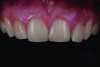
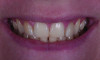
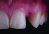
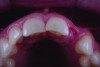
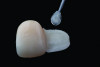
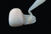
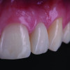
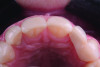
Missing teeth in the anterior region cause severe functional and, even more so, esthetic concerns. Among the main causes of missing teeth is hypodontia, the developmental absence of one or more permanent teeth. It affects roughly 2.5% to 6.9% of the population, with the maxillary lateral incisor as the most frequently absent anterior tooth.1 For clinicians, available treatment options include autotransplantation of deciduous or permanent teeth, orthodontic space closure, implant-supported restorations, cantilever resin-bonded fixed dental prostheses (RBFDPs), and conventional fixed dental prostheses (FDPs).2,3 Figure 1 and Figure 2 depict a patient with a retained deciduous maxillary left lateral incisor, which had to be removed due high mobility and advanced root resorption.
Treatment Options
When the adjacent teeth are completely intact, the use of a conventional fixed dental prothesis is usually considered overtreatment due to the significant amount of tooth structure loss that may occur during full-coverage crown preparation. Implant therapy is frequently the treatment of choice, however it is not always the most appropriate one. Patient age, insufficient space between adjacent teeth or roots, compromised hard- and soft-tissue quality, financial constraints, and patient preference are all factors that may point toward employing an alternative approach.4 This is especially true for adolescent patients and younger adults, in whom early implant placement may cause significant esthetic challenges over time due to continuous skeletal growth of the maxilla and movement of adjacent teeth, which may continue into the second and third decades of life or, in some cases, even longer.5
RBFDPs offer a minimally invasive, time- and cost-effective alternative to implant therapy, removables, and conventional crown-retained prostheses, providing excellent outcomes in durability, esthetics, function, and patient satisfaction.6 These fixed prostheses are particularly well suited for replacing lateral incisors when the adjacent abutment teeth are sound and where the pontic spaces are small. Numerous studies and systematic reviews have demonstrated very high success rates of all-ceramic RBFDPs,6-12 with recent findings reporting 98% survival of zirconia RBFDPs after 15 years.12 In addition, while biological and technical failures associated with implant-supported restorations and conventional FDPs can be catastrophic, complications with RBFDPs are usually not severe. While infrequent, the most common complication is debonding, and a debonded restoration typically can simply be rebonded rather than having to be remade. With the use of RBFDPs, more invasive options can be postponed and generally remain available later. Being able to delay more invasive tooth preparations and surgical interventions affords patients the opportunity to retain their natural teeth longer.
Ideal conditions for RBFDPs include caries-free, well-aligned abutment teeth that provide sufficient enamel for bonding. In addition, good oral hygiene, patient compliance, and the absence of deep bite, parafunctional habits, and severe periodontitis are desirable for long-term success.10
Ridge Augmentation
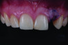
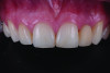
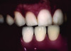
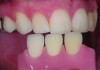
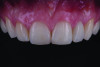
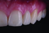
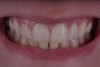
Alveolar ridge deformities in edentulous spaces or post-extraction can complicate esthetic rehabilitation. Conventional restorative solutions, such as extending the intaglio portion of the pontic, often create esthetic and functional problems, including disproportionate restoration dimensions, loss of papillae, open interdental “black triangles,” food impaction beneath the pontic, and phonetic difficulties.13 For these reasons, augmentation techniques to correct soft-tissue defects and provide functional and esthetic support are often required.14 An example is presented in Figure 3. The deciduous tooth depicted in Figure 1 and Figure 2 was extracted and a connective tissue graft was placed to improve soft-tissue volume and contour of the edentulous site. An interim removable partial denture was inserted (Figure 4) to support the tissues and shape the pontic site to create the appearance of a natural periodontal situation (Figure 5 and Figure 6).
Unlike a traditional pontic design, which emphasizes pressure-free contact over a small area, an ovate pontic applies gentle pressure to a larger area of the underlying soft tissue.15,16 This displaces tissue laterally, idealizes the form of the site, and creates an emergence profile that imitates that of a natural tooth. When the alveolar ridge is appropriately pretreated beforehand, this design has been shown to yield highly esthetic results.15 The modified ovate pontic design features a flatter contour with reduced labiolingual thickness compared with a traditional design, allowing excellent esthetics and a more precise replication of the natural tooth emergence profile.16 In addition, this technique improves access for cleaning and supports a healthy free gingival margin, while minimizing “black triangles.”17
A recent clinical observational study confirmed excellent long-term soft-tissue stability 10 years after replacing missing anterior teeth with cantilever zirconia ceramic RBFDPs.18 The authors concluded that soft tissues at pontic sites remained stable over the 10-year period with a tissue gain at the mucosal margin.
RBFDP Preparation
A key aspect for long-term success of all-ceramic RBFDPs is the single-retainer cantilever design, which offers a significantly better performance than the two-retainer design traditionally used for metal-based RBFDPs.10,19 Selecting the preferred abutment tooth follows a simple formula: the one with the largest enamel surface on the palatal aspect and the least amount of function should be chosen.
The extent of preparation of the abutment tooth depends on the available interocclusal space, overjet, and overbite. The main rule is to only prepare the tooth where space is needed and preserve as much enamel as possible. The retainer wing of the zirconia RBFDP can be as thin as 0.7 mm, which is, therefore, the minimum amount of necessary space.4 A light chamfer should be created at the gingival crest, and a margin positioned 2 mm to 3 mm from the incisal aspect. When these components are connected with proximal preparations to maximize the bonding area, a “window” to accommodate the retainer wing is created.
Finally, a small central depression facilitates accurate positioning of the definitive RBFDP during final insertion. With the proper bonding protocol, the preparation does not need any retentive elements like pins or parallel walls. In fact, to the contrary, although retentive elements may mechanically retain a debonded restoration, this could give the false impression of success while marginal leakage may be causing recurrent caries if the adhesive seal has failed.
Zirconia RBFDP
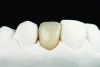
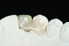
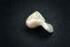
A conventional or digital impression is obtained and the shade of the anticipated restoration is communicated with the dental laboratory (Figure 7 and Figure 8). Figure 9 and Figure 10 demonstrate the RBFDP on the model. The restoration in this case was milled from 3 mol% yttria-stabilized tetragonal zirconia polycrystal (3Y-TZP), with the labial surface veneered with feldspathic ceramic. Minimal framework dimensions must be verified, with a connector width of 2 mm, connector height of 3 mm, and a retainer wing measuring a minimum thickness of 0.7 mm (Figure 11).4 These dimensions may vary depending on the type of zirconia used.
While silica-based ceramics may also be used for RBFDPs, their lower fracture strength requires significantly larger connector and retainer wing dimensions, making them unsuitable in many cases. A common misunderstanding centers around the argument that silica-based ceramics are preferred over zirconia for RBFDPs due to better “bondability.” This argument, however, overlooks the fact that tooth–ceramic bonding involves two distinctly different bonding interfaces: one to the tooth and one to the ceramic. When the bond strength to the ceramic is at least as high as the one to the tooth, the bonding interface to the tooth becomes the weak link and, therefore, the more likely cause of failure than the one at the ceramic interface. Bond strengths to dentin and enamel are typically lower than those achieved to zirconia with the proper bonding technique. This is especially true when rubber dam is not used. A clinical in situ study found 50% lower bond strength to enamel without rubber dam than with rubber dam because of the intraoral humidity and temperature changes during exhaling and inhaling.20 Therefore, better “bondability” to the ceramic has little relevance to the clinical success of the restoration as long as its bond strength is as good as or better than the one to the tooth.
The APC Zirconia Bonding Concept
RBFDPs rely on proper and long-term durable bond strengths. Conventional etching–silane treatment applied to bond to silica-based ceramics is ineffective for bonding to zirconia, especially in the long term.21 Achieving long-term durable bonding to zirconia ceramics in humid, high-stress environments is best accomplished by combining airborne-particle abrasion at moderate pressure with the application of 10-methacryloyloxydecyl dihydrogen phosphate (10-MDP)–containing primers and resin-based composite luting agents, as demonstrated in numerous studies over the past two decades.21-24 This proven protocol became popular under the term “APC concept,” comprising: (A) airborne-particle abrasion, (P) a special MDP zirconia primer, and (C) a composite resin cement (Figure 12 through Figure 14).25 The often-cited “roughening effect” of airborne-particle abrasion on zirconia is in fact only minimal.22 The main reason for improving bond strength is decontamination and a significant increase in surface free energy of the zirconia bonding surface.26 It is, therefore, critical to conduct the bonding procedure, including airborne-particle abrasion, after intraoral try-in and to apply the ceramic primer immediately after. The longer the time between airborne-particle abrasion and primer application, the lower the bond strength.26 Also, rinsing or cleaning steps are not needed after airborne-particle abrasion; loose particles can simply be removed with oil-free air flow.
In the event of the laboratory performing the airborne-particle abrasion and/or contamination before primer application, special ceramic cleaners (eg, Katana™ Cleaner, Kuraray Noritake; ZirClean®, BISCO; or Ivoclean®, Ivoclar) can be used to successfully decontaminate the zirconia bonding surface and re-establish adequate bond strength.27
Despite the proven success of the APC concept, efforts to identify alternative solutions are well underway. These include special etching solutions or silica layers applied in the laboratory. While some laboratory studies demonstrate their effectiveness, most of these approaches have only limited clinical relevance since they employ only short-term bond strength data.28 To date, APC remains the zirconia bonding protocol with the strongest support in laboratory studies and the only one proven in long-term clinical studies over 15 years.18,21
To ensure best possible bonds in the case depicted, the enamel was etched with 37% phosphoric acid for 30 seconds and a bonding agent was applied. The intaglio surface of the RBFDP retainer was airborne-particle-abraded (50-µm aluminum oxide [Al2O3] particles at 1 bar pressure). A universal ceramic primer containing functional phosphate monomers was applied and allowed to dry for about 60 seconds. The RBFDP was inserted with a resin cement. Intra- and extraoral photographs of the final restorations taken after completion of treatment are shown in Figure 15 through Figure 19.
Summary
Anterior zirconia cantilever RBFDPs have shown excellent long-term clinical performance, with survival rates of 98% after 15 years.12 The clinical success of RBFDPs depends on several interrelated factors: appropriate case selection, prosthesis design, ceramic material, bonding technique, operator skill, and follow-up care. Compared with implant therapy, RBFDPs offer the advantage of being a viable option for younger patients with potential for continued growth and in cases with limited space (<7 mm), where implant placement may be challenging. The presence of caries-free, well-aligned abutment teeth that provide sufficient enamel for bonding allows for ideal conditions for RBFDPs. In addition, good oral hygiene, patient compliance, and the absence of parafunctional habits and periodontitis are desirable factors for long-term success.10
Conclusion
Cantilever RBFDPs combined with soft-tissue augmentation represent an effective and minimally invasive approach for replacing missing anterior teeth, capable of restoring pink and white esthetics and serving as a viable alternative to implant therapy.
ABOUT THE AUTHORS
Tony Rotondo, BDSc
Private Practice, Brisbane, Australia
Szabi Hant, MDT
Private Practice, Perth, Australia
Lea S. Prott, DMD
Prosthodontist and Senior Consultant, Department of Prosthodontics, Medical Faculty and University Hospital Düsseldorf, Heinrich-Heine-University, Düsseldorf, Germany; Adjunct Professor, Department of Preventive and Restorative Sciences, University of Pennsylvania, School of Dental Medicine, Philadelphia, Pennsylvania
Markus B. Blatz, DMD, PhD
Professor of Restorative Dentistry, Chair, Department of Preventive and Restorative Sciences, and Assistant Dean, Digital Innovation and Professional Development, University of Pennsylvania, School of Dental Medicine, Philadelphia, Pennsylvania
Queries to the author regarding this course may be submitted to authorqueries@conexiant.com.
REFERENCES
1. Rakhshan V. Congenitally missing teeth (hypodontia): a review of the literature concerning the etiology, prevalence, risk factors, patterns and treatment. Dent Res J (Isfahan). 2015;12(1):1-13.
2. Kern M, Passia N, Sasse M, Yazigi C. Ten-year outcome of zirconia ceramic cantilever resin-bonded fixed dental prostheses and the influence of the reasons for missing incisors. J Dent. 2017;65:51-55.
3. Terheyden H, Wüsthoff F. Occlusal rehabilitation in patients with congenitally missing teeth-dental implants, conventional prosthetics, tooth autotransplants, and preservation of deciduous teeth – a systematic review. Int J Implant Dent. 2015;1(1):30.
4. Sasse M, Kern M. All-ceramic resin-bonded fixed dental prostheses: treatment planning, clinical procedures, and outcome. Quintessence Int. 2014;45(4):291-297.
5. Mijiritsky E, Badran M, Kleinman S, et al. Continuous tooth eruption adjacent to single-implant restorations in the anterior maxilla: aetiology, mechanism and outcomes – a review of the literature. Int Dent J. 2020;70(3):155-160.
6. Mendes JM, Bentata ALG, de Sá J, Silva AS. Survival rates of anterior-region resin-bonded fixed dental prostheses: an integrative review. Eur J Dent. 2021;15(4):788-797.
7. Naenni N, Michelotti G, Lee WZ, et al. Resin-bonded fixed dental prostheses with zirconia ceramic single retainers show high survival rates and minimal tissue changes after a mean of 10 years of service. Int J Prosthodont. 2020;33(5):503-512.
8. Kern M. Fifteen-year survival of anterior all-ceramic cantilever resin-bonded fixed dental prostheses. J Dent. 2017;56:133-135.
9. Habibzadeh S, Khamisi F, Mosaddad SA, et al. Full-ceramic resin-bonded fixed dental prostheses: a systematic review. J Appl Biomater Funct Mater. 2024;22:22808000241250118.
10. Al-Bermani ASA, Quigley NP, Ha WN. Do zirconia single-retainer resin-bonded fixed dental prostheses present a viable treatment option for the replacement of missing anterior teeth? A systematic review and meta-analysis. J Prosthet Dent. 2023;130(4):533-542.
11. Alraheam IA, Ngoc CN, Wiesen CA, Donovan TE. Five-year success rate of resin-bonded fixed partial dentures: a systematic review. J Esthet Restor Dent. 2019;31(1):40-50.
12. Kern M, Türp L, Yazigi C. Long-term outcome of anterior cantilever zirconia ceramic resin-bonded fixed dental prostheses: influence of the pontic location. J Prosthet Dent. 2025;133(4):1017-1023.
13. Zucchelli G, Mazzotti C, Bentivogli V, et al. The connective tissue platform technique for soft tissue augmentation. Int J Periodontics Restorative Dent. 2012;32(6):665-675.
14. Agarwal A, Gupta ND. Alveolar ridge augmentation by connective tissue grafting using a pouch method and modified connective tissue technique: a prospective study. Dent Res J (Isfahan). 2015;12(6):548-553.
15. Edelhoff D, Spiekermann H, Yildirim M. A review of esthetic pontic design options. Quintessence Int. 2002;33(10):736-746.
16. Gomez-Meda R, Esquivel J. Perio-prosthodontic pontic site management, part I: pontic designs and their current applications. J Esthet Restor Dent. 2023;35(4):609-620.
17. Kaufman Z, Paranhos KS. Digitally designed ovate pontic as a predictable procedure to improve accuracy, hygiene, esthetics. Compend Contin Educ Dent. 2022;43(4):226-230.
18. Türp L, Schlothauer T, Yazigi C, Kern M. Soft tissue changes at the pontic sites of cantilever zirconia ceramic resin-bonded fixed dental protheses in the esthetic zone: a clinical observational study. J Esthet Restor Dent. 2025;37(4):969-976.
19. Wei YR, Wang XD, Zhang Q, et al. Clinical performance of anterior resin-bonded fixed dental prostheses with different framework designs: a systematic review and meta-analysis. J Dent. 2016;47:1-7.
20. Falacho RI, Melo EA, Marques JA, et al. Clinical in-situ evaluation of the effect of rubber dam isolation on bond strength to enamel. J Esthet Restor Dent. 2023;35(1):48-55.
21. Blatz MB, Vonderheide M, Conejo J. The effect of resin bonding on long-term success of high-strength ceramics. J Dent Res. 2018;97(2):132-139.
22. Alammar A, Blatz MB. The resin bond to high-translucent zirconia – a systematic review. J Esthet Restor Dent. 2022;34(1):117-135.
23. Quigley NP, Loo DSS, Choy C, Ha WN. Clinical efficacy of methods for bonding to zirconia: a systematic review. J Prosthet Dent. 2021;125(2):231-240.
24. Al-Amari AS, Saleh MS, Albadah AA, et al. A comprehensive review of techniques for enhancing zirconia bond strength: current approaches and emerging innovations. Cureus. 2024;16(10):e70893.
25. Blatz MB, Alvarez M, Sawyer K, Brindis M. How to bond zirconia: the APC concept. Compend Contin Educ Dent. 2016;37(9):611-617.
26. Al-Akhali M, Al-Dobaei E, Wille S, et al. Influence of elapsed time between airborne-particle abrasion and bonding to zirconia bond strength. Dent Mater. 2021;37(3):516-522.
27. Al-Akhali M, Al-Dobaei E, Samran A, et al. Influence of cleaning methods on zirconia ceramic bonding with long-elapsed time post-airborne-particle abrasion. J Prosthodont Res. 2025;69(4):595-602.
28. D’Alessandro C, Josic U, Mazzitelli C, et al. Is zirconia surface etching a viable alternative to airborne particle abrasion? A systematic review and meta-analysis of in vitro studies. J Dent. 2024;151:105394.