You must be signed in to read the rest of this article.
Registration on CDEWorld is free. You may also login to CDEWorld with your DentalAegis.com account.
Malocclusion is highly prevalent worldwide, affecting approximately up to 75% of the global population, and nearly one in three individuals presents with a degree of malocclusion severe enough to warrant orthodontic intervention.1,2 Malocclusion also has wide-reaching impacts on esthetics, function, and patients’ overall well-being.1,3
Orthodontic therapy is a common dental treatment for children and adults. In 2024 more than six million patients underwent active orthodontic therapy in the United States, with average treatment durations ranging from 18 to 24 months.4,5 Despite its benefits, patients and dental healthcare professionals frequently cite issues such as prolonged treatment time, pain and discomfort, risk of root resorption, concerns about gingival recession, and esthetic or functional limitations as primary drawbacks and impediments to orthodontics, which underscores the demand for approaches that improve efficiency and outcomes.6 These challenges emphasize the growing clinical relevance of innovations that safely expand the limits of conventional orthodontics.
While traditional orthodontic tooth movement through fixed or removable appliances has been used to address many forms of malocclusion, it has limitations. Such limitations include anatomical restrictions of tooth movement and arch expansion as well as the methodical speed at which the tooth movement occurs. For severe cases of malocclusion, surgical interventions, such as orthognathic surgery, distraction osteogenesis, and/or corticotomy-assisted tooth movements in conjunction with the use of orthodontic appliances can enhance outcomes and accelerate tooth movement.
Surgically facilitated orthodontic therapy (SFOT) is an interdisciplinary treatment approach designed to augment orthodontic tooth movement and periodontal phenotype. SFOT has several advantages over traditional orthodontic care alone, including: (1) decreased time required for orthodontic tooth movement, (2) expansion of the envelope of feasible orthodontic movement, and (3) the ability to modify periodontal phenotype through surgical access and the potential for simultaneous hard- and/or soft-tissue grafting. Cone-beam computed tomography (CBCT), clinical assessment, photographic analysis, and intraoral scanning (IOS) are carefully used in SFOT to plan both surgical and orthodontic outcomes. SFOT requires interdisciplinary collaboration, communication, and a team approach. This article reviews critical steps in diagnosis, treatment planning, and surgical and orthodontic execution of SFOT to treat malocclusion and achieve optimal clinical and esthetic outcomes.
Background
As noted earlier, around three-fourths of individuals worldwide present with some degree of malocclusion, ranging from mild to severe forms.1,2 Malocclusion has been associated with impaired masticatory efficiency, phonetic difficulties, esthetic compromise, unfavorable airway dimensions, and reduced ability to maintain effective oral hygiene, all of which can negatively impact oral and systemic health as well as quality of life.3 Achieving optimal orthodontic outcomes, however, may be limited by biologic constraints of the dentoalveolar envelope, patient acceptance of orthognathic surgery to achieve ideal orthodontic alignment, and concerns regarding prolonged treatment duration. SFOT has emerged as an approach within interdisciplinary dentofacial therapy, enabling orthodontic tooth movement in patients whose dentoalveolar anatomy cannot safely accommodate traditional orthodontics.7
SFOT was conceived as an outgrowth of corticotomy-assisted orthodontics. It was first described by Kole and later refined by Wilcko et al through the recognition of the impact of regional acceleratory phenomenon (RAP) on orthodontic tooth movement.8 RAP enhances bone turnover and accelerates tooth movement.8 Modern SFOT integrates dentoalveolar decortication and bone and/or soft-tissue augmentation to create a bone matrix undergoing active remodeling that allows orthodontic movements to proceed more predictably and safely.9
In addition to providing enhanced orthodontic tooth movement speed and improved final outcomes, SFOT may also be indicated at sites where hard- and/or soft-tissue augmentation are anticipated to be required to avoid bony dehiscence, fenestration, and/or soft-tissue recession after completion of orthodontic realignment. Such indications may include sites presenting with thin alveolar bone or periodontal phenotype and pre-existing dehiscence or fenestration where orthodontic tooth movement is anticipated to increase the risk of gingival recession and/or hard-tissue deficiencies.10 CBCT imaging can be used to assess pre-orthodontic tooth position and bony architecture. In most patients, facial bone thickness on incisor teeth has been found to be less than 1 mm in CBCT analysis, highlighting the importance of assessing dentoalveolar phenotypes and the anatomical limitations of tooth positioning after orthodontic tooth movement prior to initiating orthodontic therapy.9 Assessment of periodontal phenotype prior to initiation of orthodontic tooth movement and utilization of computer-aided modeling to predict final tooth and hard/soft-tissue positions after ideal orthodontic alignment can allow for more comprehensive interdisciplinary planning and identify malocclusion cases that may benefit from SFOT before initiation of tooth movement.9
SFOT has been identified as advantageous in cases requiring expansion of the dentoalveolar housing or enhanced soft- and/or hard-tissue phenotype. Rationale, indications, and contraindications for SFOT are described in Table 1. SFOT has been shown to demonstrate increased long-term stability of orthodontic decompensation, particularly in cases involving crowding, transverse deficiencies, and retroclined incisors.7 When combined with hard- and soft-tissue augmentation, SFOT can enable tooth movement beyond traditional anatomic confines, reducing the risk of dehiscence, fenestration, and gingival recession while supporting long-term periodontal stability.8 Additionally, SFOT may reduce orthodontic treatment time through RAP induction and, further, may allow clinicians to avoid extractions or mitigate the esthetic and functional drawbacks of retractive orthodontics on facial contours and the airway.10 Through engagement of an interdisciplinary treatment team, SFOT can facilitate orthodontic treatment by improving dentoalveolar bone support, expand anatomical limits of traditional orthodontic tooth movement, and enhance ideal final tooth position with skeletal and soft-tissue structures to achieve predictable, long-term esthetic results.9
Diagnostic Considerations
A comprehensive diagnostic evaluation is essential before initiating SFOT, as orthodontic tooth movement must respect both the periodontal phenotype and the anatomic boundaries of the dentoalveolar complex.8 The periodontal phenotype is defined as the combined characteristics of gingival phenotype and bone morphotype; it plays a critical role in determining the limitations and long-term periodontal impacts of orthodontic tooth movement.11 Thin periodontal phenotype is characterized by decreased gingival thickness, narrow keratinized gingiva, and thin facial bone. Sites with thin periodontal phenotype are more susceptible to development or worsening of gingival recession, dehiscence, and fenestration under orthodontic forces.12 Clinical and CBCT evaluations are essential to assess soft-tissue quality and quantity, facial bone thickness, and existing recession and bony defects. Careful assessment can ensure that planned movements do not violate the alveolar housing and predispose patients to future gingival recession.
When orthodontic movements such as proclination, expansion, or decompensation exceed the anatomic envelope, patients with a thin periodontal phenotype are at higher risk for periodontal attachment loss.13 In such instances, SFOT may provide significant benefit for the stability of orthodontic outcomes and long-term periodontal health. Through the utilization and integration of CBCT and IOS technologies, clinicians can model planned orthodontic tooth movements and clinical outcomes, enhancing the predictability of final treatment outcomes and highlighting cases in which adjunctive phenotype modification and/or SFOT are indicated.14 Three-dimensional planning allows interdisciplinary teams to evaluate dentoalveolar, alveoloskeletal, and skeletal relationships simultaneously, improving treatment predictability and minimizing adverse periodontal outcomes. These tools guide decision-making regarding the need for SFOT, the extent of augmentation, and the precise anatomical regions that require corticotomy or hard/soft-tissue grafting.
SFOT is indicated when planned orthodontic movements are expected to exceed the native dentoalveolar housing, such as in cases of crowding, transverse deficiencies, or severe dental compensations.14 Patients with thin alveolar bone or those requiring high-risk decompensation movements, particularly mandibular incisor proclination, may benefit from SFOT to reduce the risk of iatrogenic dehiscence or recession. Decortications to employ RAP can accelerate tooth movement and expand the scope of orthodontic therapy for borderline orthognathic or extraction cases. Systematic reviews demonstrate that decortications used in SFOT can modestly accelerate tooth movement, supporting its use when combined with phenotype modification.15 SFOT can be highly effective for patients with dental crowding or malalignment, largely because of the accelerated tooth movement produced by RAP. SFOT can reduce treatment time by nearly half compared with conventional orthodontics.16,17
Phenotype modification may be required before, during, and/or following orthodontic tooth movement.12 The American Academy of Periodontology best evidence consensus recommends surgical intervention at sites with width of keratinized tissue of less than 2 mm, marginal tissue thickness less than 1 mm, or bone thickness less than 0.35 mm prior to tooth movement.12 Mucogingival deformities, including minimal keratinized tissue width and tissue thickness, are best treated with soft-tissue grafting procedures, namely subepithelial connective tissue grafting, which is considered the gold standard.18 Further, Wilcko and Dibart have shown hard-tissue augmentation combined with decortications to accelerate tooth movement through RAP, forming the foundation of SFOT.16,19-21 Overall, phenotype modification therapy can convert a thin, vulnerable periodontal phenotype into a more stable one, reducing risk of soft-tissue recession and bony dehiscence and improving the predictability of orthodontic outcomes. Simultaneous grafting combined with SFOT can enhance long-term periodontal and mucogingival support, improving tooth stability and reducing the risk of recession often seen after traditional orthodontic treatment.22
Surgical Considerations
Flap and incision design in SFOT are closely guided by the planned orthodontic and surgical objectives. Flap design plays a critical role in the predictability of buccal plate augmentation, access for decortication, and ideal soft-tissue grafting outcomes. A commonly used approach involves a full-thickness sulcular incision with vertical releasing incisions, providing adequate access for decortications while allowing tension-free primary closure after hard-tissue augmentation following superficial periosteal release.23 Flap elevation beyond planned decortication sites allows stable primary closure. Piezocision offers a minimally invasive, flapless approach to decortication; however, it does not permit hard- or soft-tissue grafting and may increase the risk of orthodontic force–related root resorption.21
RAP is a surgically induced, temporary surge in localized bone turnover characterized by intensified remodeling, increased vascularity, and reduced mineral density around decortication sites. This heightened biologic activity accelerates both osteoclastic and osteoblastic processes, creating an optimal environment for faster and safer orthodontic tooth movement within the alveolar housing. RAP peaks within the first several weeks post-surgery and gradually tapers over a 3- to 4-month period, making coordination of surgical treatment and orthodontic activation critical. To take full advantage of this biologic window, tooth movement should begin 1 to 2 weeks after surgery20; delaying activation risks diminishing the RAP effect, reducing the anticipated acceleration, and potentially affecting graft integration.
Decortications involve cutting through the cortical plate and into marrow space to stimulate localized bone remodeling response, ie, RAP. In animal investigations, sites treated with decortication showed significantly higher osteoclastic activity at 3 days and increased expression of pro-inflammatory mediators associated with bone turnover, including transforming growth factor (TGF)-β1, vascular endothelial growth factor (VEGF), and osteocalcin, by 21 days.19,24 These findings suggest that decortications stimulate accelerated bone turnover and improved regenerative activity.16,25 Cohen et al demonstrated that deeper penetration into medullary bone intensifies RAP effect, promoting faster remodeling and enhanced graft integration.26 Their study indicates that initial surgical intervention should take into account planned tooth movements to allow for appropriate decortication depth and location. Decortications should be tailored to support blood supply, graft turnover (if applicable), and predictable bone formation.
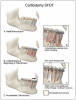
Techniques to induce RAP are commonly divided into two categories: (1) procedures that rely primarily on decortication and (2) those that involve single or multiple tooth osteotomies. Decortication selectively perforates or modifies the cortical bone to the level of the medullary bone while preserving medullary continuity, whereas an osteotomy involves complete cuts through both cortical and medullary bone, typically creating a mobilized bone segment (Figure 1). By inducing RAP, decortication effectively enhances conventional orthodontic therapy by increasing the biologic plasticity of the dentoalveolar complex. This heightened responsiveness facilitates more efficient tooth movement, particularly rotational and tipping movements, and supports moderate arch development. Following healing, orthodontic outcomes achieved with decortication-assisted approaches appear highly stable, likely because of reduced tissue memory as compared with traditional orthodontic tooth movement.
Piezoelectric surgery was introduced as a less traumatic alternative to rotary instrumentation, selectively cutting mineralized tissue while preserving soft tissues, blood supply, and osteocyte viability, thereby supporting more favorable bone healing.27 Although a split-mouth animal study reported faster tooth movement with rotary burs, likely due to greater trauma-induced RAP activation, the clinical significance of this acceleration remains uncertain given potential adverse effects on bone vitality and long-term remodeling.28
Limited evidence exists that quantifies the ideal amount of hard-tissue augmentation; however, when planning it, the existing periodontal phenotype direction and the magnitude of planned tooth movement should be accounted for. Close interdisciplinary collaboration is essential to determine these movements and guide tissue augmentation needs prior to surgery. Factors influencing graft volume include CBCT accuracy in assessing buccal bone thickness, potential coronal graft loss from flap tension, expected graft resorption, healing response, and the degree of tooth tipping and/or translation. Because graft resorption of roughly one-third is common in guided bone regeneration (GBR) procedures, over-grafting the buccal plate is often recommended to compensate for future anticipated loss and ensure adequate postoperative bone thickness to allow for optimal tooth position.28
Common graft materials used in SFOT include autogenous bone, demineralized freeze-dried bone allograft, freeze-dried bone allograft, deproteinized bovine bone matrix (DBBM), or composite grafts comprised of a combination of these materials, although comparative evidence between bone replacement graft types remains limited. Because SFOT frequently involves non-contained defects, grafts must provide reliable long-term space maintenance. DBBM demonstrates slower resorption, superior space stability, and histologic integration with newly formed bone while still permitting orthodontic tooth movement, making its use in grafting advantageous compared to faster-resorbing grafts alone for buccal augmentation.27 Although direct evidence for biologics in SFOT is limited, their proven benefits in guided bone and tissue regeneration may support their use in this context.22 Commonly used biologics include recombinant human platelet-derived growth factor-BB, enamel matrix derivative, and autologous blood products such as platelet-rich plasma and platelet-rich fibrin.29,30
Barrier membranes are generally recommended in GBR to provide wound stability and maintain space. Because buccal plate grafting lacks natural containment once the flap is elevated, a membrane is often critical for optimal outcomes.22 Although specific barrier membrane superiority cannot be identified in the current literature, use of acellular dermal matrix (ADM) may be desirable clinically to allow for simultaneous barrier function and soft-tissue phenotype enhancement potential. ADM, however, may carry risks related to poor hydrophilicity and lack of vasculature, including postoperative sloughing, graft exposure, and long-term shrinkage, all of which may compromise bone volume and soft-tissue outcomes.31,32 Other barrier membranes, such as cross-linked collagen membranes, may provide longer-lasting barrier function of up to 6 months and have demonstrated effective support for osseous regeneration.33
Interdisciplinary Teams for Optimal Treatment Outcomes
Interdisciplinary dentofacial therapy (IDT) requires tightly coordinated collaboration among the restorative dentist, periodontist, orthodontist, oral and maxillofacial surgeon, and other medical partners when indicated.10 The goal of IDT is to diagnose craniofacial, periodontal, and dental problems comprehensively and to plan treatment from the perspective of facial harmony, functional stability, and long-term periodontal health. Contemporary IDT recognizes that clinical signs such as crowding, compensatory incisor inclinations, thin facial bone, or worn dentition represent deeper skeletal and dentoalveolar imbalances.10 CBCT, IOS, mounted models, and digital workflows allow the team to visualize tooth-to-bone, tooth-to-jaw, and jaw-to-face relationships with unprecedented precision. This collaborative planning process enables formation of a unified, restoratively driven plan that respects biologic boundaries, anticipates the need for phenotype modification or bone augmentation, and aims to deliver esthetic and functional results that are sustainable over the patient’s lifetime.
Modern treatment planning begins with a facially prioritized philosophy in which teeth and supporting structures are ideally positioned relative to the lips, facial profile, smile dynamics, and airway volume. Instead of simply aligning teeth to fit existing bone, clinicians should evaluate how tooth position will influence facial esthetics, lip support, buccal corridors, vertical facial proportions, and long-term occlusal stability. By designing treatment from the outside-in and with the final result in mind, the team can determine whether orthodontics alone can achieve facial goals or if procedures such as SFOT, phenotype modification, and/or orthognathic surgery are needed for optimal results. This approach facilitates the achievement of desired esthetic outcomes, such as ideal incisor display, harmonious profile, and a stable smile arc, without compromising periodontal health or creating iatrogenic damage.
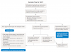
Digital workflows, particularly CBCT and IOS-based orthodontic simulation software, are central to evaluating whether a patient is an appropriate candidate for SFOT. These tools allow clinicians to visualize existing dentoalveolar bone volume, identify dehiscence or fenestration, measure the initial anatomic limits of orthodontic movement and simulate proposed tooth movements to determine whether they exceed biologic limits.10 For patients with crowding, retroclined incisors, thin facial bone, or skeletal disharmony, simulations often reveal that conventional orthodontics would place roots outside the alveolar housing, highlighting the need for SFOT-assisted decompensation. Image-based modeling allows for optimal case selection, risk assessment, and patient communication. This clarity enhances collaborative decision-making, leading to safer, highly predictable, and more stable orthodontic outcomes (Figure 2).
Clinical Outcomes
SFOT may provide significant advantages over traditional orthodontic therapy in the efficiency and predictability of orthodontic tooth movement. Further, combining phenotype modification with hard- and soft-tissue grafting can increase the available envelope of tooth movement and allow for post-orthodontic stability of hard and soft tissues. Clinical case series consistently show robust periodontal architecture and stable bone support 1-year post-SFOT, even in patients with pre-existing defects or thin periodontal phenotypes.8,10 Achieving predictable results requires careful diagnosis, collaborative interdisciplinary planning, appropriate flap and biomaterial selection, controlled and monitored orthodontic tooth movement, and proper postoperative management to appropriately address underlying anatomic risk factors and patient and clinician concerns.
SFOT can allow long-term dental health and stability through enhanced hard- and soft-tissue architecture. Bone grafting and soft-tissue augmentation performed during SFOT meaningfully increase buccal bone volume, improve gingival thickness, and help prevent post-orthodontic recession. Beyond periodontal outcomes, SFOT also contributes positively to airway and sleep physiology by enabling non-retractive orthodontic strategies. By expanding the dentoalveolar housing and allowing forward positioning of teeth and tongue posture, SFOT may help increase oral cavity volume and improve apnea-hypopnea indices in select patients, supporting a more open and physiologically favorable upper airway.34 These systemic benefits highlight SFOT’s expanding role within interdisciplinary dentofacial and airway-focused treatment planning.8,10,34
Summary and Clinical Decision-Making
SFOT expands orthodontic treatment possibilities for patients whose malocclusion exceeds conventional biologic limits, offering a multidisciplinary approach to address complex dentoalveolar, periodontal, and skeletal constraints. Successful application requires advanced surgical skill and close interdisciplinary coordination to align diagnosis, treatment sequencing, timing, and surgical protocols, allowing the treatment team to address underlying biologic limitations rather than tooth alignment alone. Integration of 3-dimensional imaging and digital treatment modeling enhances diagnostic transparency, supports biologically sound planning, and fosters shared accountability among providers, reducing the need for more invasive orthognathic interventions.
Despite these advantages, SFOT has inherent limitations and is not appropriate for all patients or clinical settings. Careful patient selection is essential, as contraindications include active periodontal or endodontic disease, insufficient attached gingiva, uncontrolled systemic conditions, compromised immune status, poor compliance, and the use of medications that impair bone metabolism; nonsteroidal anti-inflammatory drugs may further diminish treatment efficacy by attenuating the regional acceleratory phenomenon.35 SFOT does not improve clinical attachment levels and should not be viewed as a rescue technique in periodontally compromised dentitions, as pre-existing bone loss may result in pocket reduction and increased clinical crown length rather than true regeneration. Additionally, ankylosed teeth or teeth within devitalized bone cannot be predictably moved, and inadequate decortication may compromise alveolar support without eliciting a sufficient biologic response.16
Although long-term outcome data remains limited, current evidence suggests that SFOT does not increase the risk of excessive root resorption, as accelerated tooth movement is driven by transient bone demineralization rather than excessive orthodontic forces.16 Ultimately, when applied judiciously within an interdisciplinary framework, SFOT offers a biologically grounded strategy to achieve stable, patient-centered outcomes that extend beyond the pursuit of straight teeth.
ABOUT THE AUTHORS
Priscilla Sosa, DMD
Private Practice limited to Periodontology, Sarasota, Florida
Maria L. Geisinger, DDS, MS
Professor and Chair, Kent and Phoebe Endowed Professor in Periodontology, Director, Advanced Education in Periodontology, Department of Periodontology, University of Alabama at Birmingham School of Dentistry, Birmingham, Alabama; Diplomate, American Board of Periodontology
Ramzi V. Abou-Arraj, DDS, MS
Professor, Associate Dean for Academic Affairs, Department of Periodontology, University of Alabama at Birmingham School of Dentistry, Birmingham, Alabama
Queries to the author regarding this course may be submitted
to authorqueries@conexiant.com.
REFERENCES
1. Profitt WR, Fields HW Jr, Moray LJ. Prevalence of malocclusion and orthodontic treatment need in the United States: estimates from the NHANES III survey. Int J Adult Orthodon Orthognath Surg. 1998;13(2):97-106.
2. Lombardo G, Vena F, Negri P, et al. Worldwide prevalence of malocclusion in the different stages of dentition: a systematic review and meta-analysis. Eur J Paediatr Dent. 2020;21(2):115-122.
3. Bollen AM. Effects of malocclusions and orthodontics on periodontal health: evidence from a systematic review. J Dent Educ. 2008;72(8):912-918.
4. Aljehani D, Baeshen HA. Effectiveness of the American Board of Orthodontics discrepancy index in predicting treatment time. J Contemp Dent Pract. 2018;19(6):647-650.
5. American Association of Orthodontists. Member survey indicates orthodontic patient numbers at all-time high. AAO website. July 9, 2025. https://www2.aaoinfo.org/member-survey-indicates-orthodontic-patient-numbers-at-all-time-high/. Accessed April 22, 2026.
6. Tsichlaki A, O’Brien K. Do orthodontic research outcomes reflect patient values? A systematic review of randomized controlled trials involving children. Am J Orthod Dentofacial Orthop. 2014;146(3):279-285.
7. Mandelaris GA, Richman C, Kao RT. Surgical considerations and decision making in surgically facilitated orthodontic treatment/
periodontally accelerated osteogenic orthodontics. Clin Adv Periodontics. 2020;10(4):213-223.
8. Dounis T, Pitman LM. Decision making for soft and hard tissue augmentation in surgically facilitated orthodontics. Clin Adv Periodontics. 2020;10(1):38-41.
9. Mandelaris GA, DeGroot BS, Relle R, et al. Surgically facilitated orthodontic therapy: optimizing dentoalveolar bone and space appropriation for facially prioritized interdisciplinary dentofacial therapy. Compend Contin Educ Dent. 2018;39(3):146-156.
10. Mandelaris GA, Huang I, Relle R, et al. Surgically facilitated orthodontic therapy (SFOT): diagnosis and indications in interdisciplinary dentofacial therapy involving tooth movement. Clin Adv Periodontics. 2020;10(4):204-212.
11. Cortellini P, Bissada NF. Mucogingival conditions in the natural dentition: narrative review, case definitions, and diagnostic considerations. J Periodontol. 2018;89(suppl 1):S204-S213.
12. Kao RT, Curtis DA, Kim DM, et al. American Academy of Periodontology best evidence consensus statement on modifying periodontal phenotype in preparation for orthodontic and restorative treatment. J Periodontol. 2020;91(3):289-298.
13. Wang CW, Yu SH, Mandelaris GA, Wang HL. Is periodontal phenotype modification therapy beneficial for patients receiving orthodontic treatment? An American Academy of Periodontology best evidence review. J Periodontol. 2020;91(3):299-310.
14. Roblee RD, Bolding SL, Landers JM. Surgically facilitated orthodontic therapy: a new tool for optimal interdisciplinary results. Compend Contin Educ Dent. 2009;30(5):264-275.
15. Fleming PS, Fedorowicz Z, Johal A, et al. Surgical adjunctive procedures for accelerating orthodontic treatment. Cochrane Oral Health Group, ed. Cochrane Database Syst Rev. 2015;2015(6):CD010572.
16. Wilcko W, Wilcko MT. Accelerating tooth movement: the case for corticotomy-induced orthodontics. Am J Orthod Dentofacial Orthop. 2013;144(1):4-12.
17. Gil APS, Haas OL Jr, Méndez-Manjón I, et al. Alveolar corticotomies for accelerated orthodontics: a systematic review. J Craniomaxillofac Surg. 2018;46(3):438-445.
18. Barootchi S, Tavelli L, Zucchelli G, et al. Gingival phenotype modification therapies on natural teeth: a network meta analysis. J Periodontol. 2020;91(11):1386-1399.
19. Wilcko MT, Ferguson DJ, Makki L, Wilcko WM. Keratinized gingiva height increases after alveolar corticotomy and augmentation bone grafting. J Periodontol. 2015;86(10):1107-1115.
20. Wilcko MT, Wilcko WM, Pulver JJ, et al. Accelerated osteogenic orthodontics technique: a 1-stage surgically facilitated rapid orthodontic technique with alveolar augmentation. J Oral Maxillofac Surg. 2009;67(10):2149-2159.
21. Dibart S. Piezocision™: Accelerating orthodontic tooth movement while correcting hard and soft tissue deficiencies. In: Kantarci A, Will L, Yen S, eds. Tooth Movement (Frontiers of Oral Biology). Vol 18. Basel, Switzerland: S. Karger AG; 2016:102-108.
22. Stern JK, Beauchamp S, Baldock W, et al. Factors affecting predictability of buccal bone augmentation in surgically facilitated orthodontic treatment: surgical considerations. Compend Contin Educ Dent. 2020;41(8):410-418.
23. Greenwell H, Vance G, Munninger B, Johnston H. Superficial-layer split-thickness flap for maximal flap release and coronal positioning: a surgical technique. Int J Periodontics Restorative Dent. 2004;24(6):521-527.
24. Wang L, Lee W, Lei DL, et al. Tissue responses in corticotomy- and osteotomy-assisted tooth movements in rats: histology and immunostaining. Am J Orthod Dentofacial Orthop. 2009;136(6):770.e1-770.e11.
25. Baloul SS, Gerstenfeld LC, Morgan EF, et al. Mechanism of action and morphologic changes in the alveolar bone in response to selective alveolar decortication–facilitated tooth movement. Am J Orthod Dentofacial Orthop. 2011;139(4 suppl):S83-S101.
26. Cohen G, Campbell PM, Rossouw PE, Buschang PH. Effects of increased surgical trauma on rates of tooth movement and apical root resorption in foxhound dogs. Orthod Craniofac Res. 2010;13(3):179-190.
27. Araújo MG, Carmagnola D, Berglundh T, et al. Orthodontic movement in bone defects augmented with Bio‐Oss. An experimental study in dogs. J Clin Periodontol. 2001;28(1):73-80.
28. Elnayef B, Porta C, Suárez-López Del Amo F, et al. The fate of lateral ridge augmentation: a systematic review and meta-analysis. Int J Oral Maxillofac Implants. 2018;33(3):622-635.
29. Ghanaati S, Herrera-Vizcaino C, Al-Maawi S, et al. Fifteen years of platelet rich fibrin in dentistry and oromaxillofacial surgery: how high is the level of scientific evidence? J Oral Implantol. 2018;44(6):471-492.
30. Miron RJ, Zucchelli G, Pikos MA, et al. Use of platelet-rich fibrin in regenerative dentistry: a systematic review. Clin Oral Investig. 2017;21(6):1913-1927.
31. Ozturan S, Oztunc H, Evlice BK. Assessment of the soft tissue volumetric changes following acellular dermal matrix grafts with cone beam computerized tomography. Quintessence Int. 2015;46(2):171-178.
32. Harris RJ. A short‐term and long‐term comparison of root coverage with an acellular dermal matrix and a subepithelial graft. J Periodontol. 2004;75(5):734-743.
33. Zubery Y, Goldlust A, Alves A, Nir E. Ossification of a novel cross‐linked porcine collagen barrier in guided bone regeneration in dogs. J Periodontol. 2007;78(1):112-121.
34. Mandelaris GA, DeGroot BS, Relle R, et al. Surgically facilitated orthodontic therapy: optimizing dentoalveolar bone and space appropriation for facially prioritized interdisciplinary dentofacial therapy. Compend Contin Educ Dent. 2018;39(3):146-156.
35. Zimmo N, Saleh MHA, Mandelaris GA, et al. Corticotomy-accelerated orthodontics: a comprehensive review and update. Compend Contin Educ Dent. 2017;38(1):17-26.