You must be signed in to read the rest of this article.
Registration on CDEWorld is free. You may also login to CDEWorld with your DentalAegis.com account.
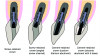
When restoring an anterior dental implant, a clinician must decide whether to use a screw- or cement-retained crown. A recent survey of US dentists reported that 80% of respondents would restore an ideally placed anterior implant with angulation through the palatal surface with a screw-retained implant crown (Figure 1, A).1 Perhaps the biggest advantage of a screw-retained crown versus a cement-retained one is the avoidance of excess cement below the crown margin. Such excess cement can lead to peri-implantitis and has been attributed to bone loss around cement-retained restorations, with 2.8% of cement-retained restorations and 0% of screw-retained restorations showing more than 2 mm of bone loss at 5 years.2 Screw-retained implant crowns also offer the advantage of retrievability.2
Clinicians face a more difficult decision if the implant is angled through the facial aspect of a proposed crown, as a screw-channel access would then be visible on the facial surface of the crown. In the case of an anterior implant angled through the facial, 55% of respondents in the aforementioned survey would use an angled screw system, and 41% would use a cement-retained crown.1 Angled screw-channel abutments have an access opening that can be angled up to about 25 degrees to the palatal but require a ball-end driver to tighten the abutment screw (Figure 1, B).
Of those respondents who chose to use a cement-retained crown, 33% would employ custom titanium abutments (Figure 1, C) while 42% would use zirconia abutments with a titanium base (Figure 1, D).1 A clinical concern with cement-retained restorations is the show-through of the titanium abutment through the crown ceramic or soft tissue. When using a zirconia abutment with a titanium base this issue is mostly negated, because the zirconia abutment shade can be selected to match the restoration or surrounding teeth preparations (Figure 1, D). With regard to soft tissue, the zirconia portion of the abutment below the soft tissue has been reported to provide a more esthetic result than titanium,3 as titanium may cause soft tissue to appear dark, especially in patients with a thin gingival biotype. Additionally, the collar of the titanium base in such a restoration can be as thin as 0.7 mm and, therefore, have no negative influence on bone stability4 while enabling the titanium base to be essentially hidden within the sulcus.
Some clinicians prefer to use custom titanium abutments rather than a zirconia abutment for cement-retained crowns (Figure 1, C) to avoid technical issues regarding the bond between the titanium base and zirconia abutment. The problem of show-through with a titanium abutment, however, is more troublesome than with a zirconia abutment. Results of the previously referenced survey revealed that cement-retained crowns would be fabricated from zirconia (42%), lithium disilicate (33%), or metal-ceramic (25%).1 Metal-based crowns would not be affected by the color of the underlying titanium, however the relatively translucent ceramic materials would be affected. The crowns could be designed to be thicker to better block the color of the titanium abutment, but their thickness would be limited by the space needed for adequate thickness of the abutment walls.
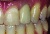
Titanium show-through of the gingival can also be an issue. Figure 2 demonstrates a titanium abutment that is visible through the soft tissue of an anterior implant restoration. Results of the aforementioned survey disclosed that 42% of clinicians place the abutment margins 1 mm subgingival and the remainder are placed 0.5 mm subgingival (33%) or equigingival (25%).1 Placing the margins deeper within the sulcus may help to hide the titanium; however, margins placed 1 mm and 3 mm subgingivally had about 12 times or 24 times more undetected excess cement, respectively, than equigingival margins.5
Anodization: Altering the Color of Titanium
One strategy to compensate for the color of a titanium abutment is to anodize the abutment. Anodization (anodic oxidation) is a method used to alter the color of titanium by creating oxide layers of different thicknesses, which results in color changes through light interference. Varying the voltage applied to the titanium affects the thickness of the oxide layer and resulting color. For example, specific voltages can create shades of gold or pink on the titanium surface. Compared to untreated titanium, anodized abutments showed increased roughness and decreased contact angle; however, cell morphology, proliferation, and viability of gingival fibroblasts on anodized titanium were comparable to untreated titanium.6 Anodized titanium abutments have been shown to produce histological and clinical outcomes similar to untreated titanium.7
Several studies have reported that gold anodization of titanium abutments can be used to more effectively match the color of overlying lithium-disilicate restorations to surrounding teeth. In one study, 1 mm, 1.5 mm, and 2 mm thick lithium-disilicate specimens were bonded to gold anodized and unanodized titanium blocks. Their color was compared to a 5 mm thick block of lithium disilicate. Gold anodization allowed better color match of lithium disilicate at all thicknesses.8 Another study examined lithium-disilicate specimens (between 1 mm and 2.5 mm thickness) against zirconia, gold anodized titanium, and unanodized titanium. The lithium disilicate against gold anodized titanium matched the lithium disilicate against an A2 dentin shade background better than any other background.9
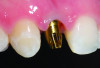
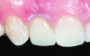
Figure 3 and Figure 4 show a gold anodized abutment and an overlying lithium-disilicate restoration, respectively. Zirconia is a relatively opaque ceramic, and studies are less clear as to the benefit of the use of gold anodized abutments under zirconia implant crowns. One study of 0.7 mm to 1.1 mm thick zirconia specimens reported that gold anodization of a titanium background did not improve the color match to a zirconia crown against a zirconia background.10 On the other hand, another study reported that the color change of 0.5 mm thick zirconia specimens against titanium backgrounds could be improved by gold anodization.11
Anodizing titanium abutments to a pink color can mask the color of titanium abutments through soft tissue. In a study, four different abutments were fabricated for each participant: gold anodized titanium, pink anodized titanium, unanodized titanium, and zirconia. After insertion of the abutments and their corresponding crowns, the color of the peri-implant soft tissue and the natural gingiva on the opposite side were measured using a spectrophotometer. The study revealed that the mean color difference from lowest to highest was ranked as zirconia, pink anodized titanium, gold anodized titanium, and unanodized titanium.12
Another study involved 25 patients with a missing maxillary tooth in the esthetic region and included 25 crowns on gray titanium, pink anodized titanium, and hybrid zirconia abutments. Color measurements of the soft tissue of the implant site and contralateral tooth were compared. The results showed that the hybrid zirconia abutments produced the least color difference, followed closely by the pink anodized titanium abutments. Gray titanium abutments had the greatest color difference.13 In yet another study of 40 patients, it was determined that changing abutments of provisional anterior crowns from gray to pink significantly improved the color of the peri-implant mucosa, making it more closely match the natural gingiva. This color change was particularly noticeable in patients with thin gingival biotypes.14 In summary, pink anodization of abutments can allow for a better match of surrounding soft tissue than unanodized titanium but not as good a match as zirconia abutments.
Pink anodization of abutments to better match soft tissue does have a negative effect on the color match of ceramic crowns. When using lithium-disilicate crowns, pink anodized abutments created greater color change from a dentin background than the use of gold or unanodized titanium abutments.9 A similar trend was seen for 0.5 mm thick zirconia specimens.11
The Process to Anodize Titanium Abutments
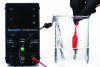
The process of anodizing titanium is fairly simple and requires only a source of direct current and a container with an electrolytic solution (Figure 5). The source of the direct current may be a DC power supply or a series of batteries. A low current of a maximum of 3A should be used.15 Different electrolytic solutions have been suggested, including a trisodium phosphate (TSP) heavy-duty cleaner (1 g TSP to 500 mL water) and 1M phosphoric acid solution. The use of different electrolytic solutions will affect the voltage required to obtain a desired color.6,15,16
To anodize an implant abutment, the negative terminal of the power supply is attached to a 3 cm x 6 cm piece of aluminum foil, which is submerged into the electrolytic solution (in an approximately 250 mL container). The positive terminal is connected to the clean titanium abutment, which is then submerged into the electrolytic solution. Complete anodization occurs in 10 to 20 seconds. Even small voltages can cause electrical shock; therefore, the terminal leads (negative and positive) should not be touched when the power supply is on, and appropriate insulation, such as rubber gloves, must be worn throughout the process.15
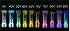
When using a TSP electrolytic solution, gold hue is achieved from 60V to 65V and pink hue is achieved from 70V to 80V.16 A color chart based on a TSP electrolytic solution is presented in Figure 6. When using 1M phosphoric acid solution, gold hues are achieved at 50V to 60V and pink hues at 65V to 70V.6
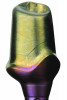
An implant part can always be subjected to an additional soaking at a higher voltage to further change the color of the part; however, dipping at a lower voltage will not return the part to an original color. Therefore, it is prudent to start at a low voltage and observe the achieved color change when first attempting this procedure. An implant part can also attain more than one color by being selectively dipped in the solution. For example, an abutment could be dipped in solution at 60V to 65V to obtain a gold hue, and then the subgingival component of the abutment could be dipped in solution at 75V to 80V to gain a pink hue (Figure 7).15
Evaluation of Color Change: Lithium Disilicate and Zirconia Against Anodized Titanium
Previous studies have suggested that gold anodization of titanium backgrounds may improve the color match of lithium-disilicate and zirconia crowns and that pink anodization may worsen the color match.8,9,11 The present investigation aimed to compare the color of zirconia and lithium-disilicate specimens at varying thicknesses (0.5 mm to 2 mm) against various shades of titanium to a reference of dentin shade composite. The goal of the investigation was to determine: (1) if the hue of titanium affected the final color of either of the ceramic materials, and (2) what thickness of each ceramic over different hues of titanium would allow adequate color match with the same ceramic over dentin.
Titanium blanks (Ti-6Al-4V, BioHorizons) were sectioned into three 6 mm blocks and wet-polished to 1,200 grit silicon carbide paper. The titanium blocks were anodized using the process described in the previous section. Using a TSP electrolytic solution, one block was anodized gold (60V) and one block was anodized pink (80V). An A2 dentin shade composite (Filtek™ Supreme A2D, 3M Oral Care) was shaped into a 6 mm thick block and cured.
Specimens of lithium disilicate (IPS e.max® LT A2, Ivoclar) and 3 mol% yttria stabilized zirconia (ZirCAD HT A2, Ivoclar) were sectioned into tiles using a sectioning saw. The ceramics were crystallized or sintered according to their manufacturer's directions and then polished to 1,200 grit silicon carbide paper. The final dimensions of the tiles were confirmed with the use of a caliper to be 0.5 mm, 0.8 mm, 1 mm, 1.2 mm, 1.5 mm, 1.8 mm, and 2 mm.
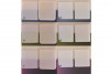
The ceramic tiles were placed over the titanium or dentin shade composite blocks without any intermediary medium (ie, cement). For each combination of ceramic tile (type of ceramic and thickness) and titanium (gold anodized, pink anodized, unanodized) or dentin shade composite block, L*, a*, and b* color values were recorded with a spectrophotometer. The difference in L*, a*, and b* color between each ceramic tile against the titanium blocks and their color against the dentin shade composite block was compared using the ΔE2000 formula. Representative tiles overlaying titanium and dentin shade composite blocks are shown in Figure 8.
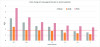
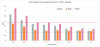
ΔE2000 represents the color difference between two objects, where a higher ΔE2000 value indicates a greater difference in color, making the variation more perceptible to the human eye. In dentistry, ΔE2000 plays an important role in assessing both the perceptibility and acceptability of color differences between dental restorations and natural teeth. The perceptibility threshold refers to the smallest color variation noticeable to the human eye. A study reported an average ΔE2000 perceptibility threshold of 0.81 (ranging from 0.34 to 1.28) that was detectable by 50% of observers. On the other hand, the acceptability threshold refers to the largest color difference that is still considered clinically acceptable. This threshold is a ΔE2000 value of 1.77 (ranging from 1.23 to 2.37), with 50% of observers considering it acceptable.16
The results of this investigation are presented in Figure 9 and Figure 10 along with demarcations for the threshold of clinical perceptibility and acceptability. These results confirm the results of previous studies that gold anodization of titanium improves color match and pink anodization worsens color match for both lithium disilicate and zirconia. For lithium disilicate, the threshold of clinical acceptable color match with a dentin shade background could be achieved at 1 mm thickness for gold anodized titanium and 1.8 mm with unanodized titanium. This threshold could not be achieved with pink anodized titanium. For zirconia, the threshold of clinical acceptable color match with a dentin shade background could be achieved at 0.5 mm thickness for gold anodized titanium and 1 mm with unanodized titanium and pink anodized titanium.
In summary, restoring a facially angled implant with a screw-retained angled screw channel crown or a crown with a zirconia abutment may lead to debonding issues of the crown from the titanium base. The use of a custom titanium abutment may avoid this technical complication. To overcome the esthetic challenge of placing a ceramic crown on a titanium abutment, the abutment can be anodized gold (60V to 65V) and then a 1 mm lithium-disilicate or 0.5 mm zirconia restoration would be able to acceptably mask the color of the abutment. If pink anodization of the abutment (75V to 80V) is needed to mask the color of the abutment under soft tissue, 1 mm of zirconia is needed to acceptably mask the color of the abutment. Preferably, in this clinical circumstance, the abutment could be selectively anodized at the subgingival portion only.
Conclusion
Selecting the optimal restoration method for anterior implants requires careful consideration of both functional and esthetic outcomes. While screw-retained crowns offer retrievability and minimize cement-related complications, cement-retained crowns with anodized titanium or zirconia abutments provide viable alternatives. Anodization of titanium abutments presents a practical strategy to improve esthetics, with gold hues enhancing ceramic crown color matching and pink hues better blending with soft tissue. Clinicians can optimize outcomes by tailoring ceramic thickness and anodization selectively, balancing technical feasibility with esthetic demands to achieve patient-satisfying results.
Acknowledgment
The authors thank Mark Ferguson of Vulcan Custom Dental for donating the titanium blocks used in this study, and Hannah Bloom, Andrew Lisy, and Brandon Englert for their help with data collection and project inception.
About the Authors
Mohammed Hammamy, DDS
First-Year Resident, Biomaterials Residency Program, University of Alabama at Birmingham School of Dentistry, Birmingham, Alabama
Silvia Rojas-Rueda, DDS
Graduate Student, Division of Biomaterials, Department of Clinical and Community Sciences, University of Alabama at Birmingham School of Dentistry, Birmingham, Alabama
Jonathan Esquivel, DDS
Private Practice in Prosthodontics, Metairie, Louisiana
Nathaniel C. Lawson, DMD, PhD
Director, Division of Biomaterials, and Program Director of the Biomaterials Residency Program, University of Alabama at Birmingham School of Dentistry, Birmingham, Alabama; General Dentist, UAB Faculty Practice
Queries to the author regarding this course may be submitted to authorqueries@conexiant.com.
References
1. Schoenbaum TR, Papaspyridakos P, Kim YK, et al. Clinician preferences for single-unit implant restoration designs and materials: a survey of the membership of the Pacific Coast Society for Prosthodontics. J Prosthet Dent. 2024;132(6):1288-1298.
2. Sailer I, Mühlemann S, Zwahlen M, et al. Cemented and screw-retained implant reconstructions: a systematic review of the survival and complication rates. Clin Oral Implants Res. 2012;23 suppl 6:163-201.
3. Linkevicius T, Vaitelis J. The effect of zirconia or titanium as abutment material on soft peri-implant tissues: a systematic review and meta-analysis. Clin Oral Implants Res. 2015;26 suppl 11:139-147.
4. Linkevicius T, Alkimavicius J, Linkevicius R, et al. Effect of ti-base abutment gingival height on maintenance of crestal bone in thick biotype patients: a randomized clinical trial with 1-year follow-up. Int J Oral Maxillofac Implants. 2022;37(2):320-327.
5. Linkevicius T, Vindasiute E, Puisys A, et al. The influence of the cementation margin position on the amount of undetected cement. A prospective clinical study. Clin Oral Implants Res. 2013;24(1):71-76.
6. Wang T, Wang L, Lu Q, Fan Z. Changes in the esthetic, physical, and biological properties of a titanium alloy abutment treated by anodic oxidation. J Prosthet Dent. 2019;121(1):156-165.
7. Areid N, Abushahba F, Riivari S, Närhi T. Effect of TiO2 abutment coatings on peri-implant soft tissue behavior: a systematic review of in vivo studies. Int J Dent. 2024;2024:9079673.
8. Farrag KM, Bakry SI, Aly YM. Effect of yellow anodization of titanium on the shade of lithium disilicate ceramic with different thicknesses. J Prosthet Dent. 2022;128(4):793.e1-793.e6.
9. Weeranoppanant P, Palanuwech M. Effects of ceramic thickness and titanium anodization on esthetic outcomes of lithium disilicate ceramic over titanium alloys. Eur J Prosthodont Restor Dent.2023;31(1):40-49.
10. Bas BB, Cakan U. Evaluation of the effect of anodization-colored titanium abutments and zirconia substructure thickness on zirconia substructure color: an in vitro study. Niger J Clin Pract. 2022;25(12):2024-2029.
11. DeGirmenci K, Saridag S. Influence of anodized titanium abutment backgrounds on the color parameters of different zirconia materials. Am J Dent. 2021;34(1):39-43.
12. Wang T, Wang L, Lu Q, Fan Z. Influence of anodized titanium abutments on the esthetics of the peri-implant soft tissue: a clinical study. J Prosthet Dent. 2021;125(3):445-452.
13. Vazouras K, Gholami H, Margvelashvili-Malament M, et al. An esthetic evaluation of different abutment materials in the anterior maxilla: a randomized controlled clinical trial using a crossover design. J Prosthodont. 2022;31(8):673-680.
14. Bittner N, Schulze-Späte U, Silva C, et al. Comparison of peri-implant soft tissue color with the use of pink-neck vs gray implants and abutments based on soft tissue thickness: a 6-month follow-up study. Int J Prosthodont. 2020;33(1):29-38.
15. Wadhwani CP, O'Brien R, Kattadiyil MT, Chung KH. Laboratory technique for coloring titanium abutments to improve esthetics. J Prosthet Dent. 2016;115(4):409-411.
16. Paravina RD, Ghinea R, Herrera LJ, et al. Color difference thresholds in dentistry. J Esthet Restor Dent. 2015;27 suppl 1:S1-S9.