You must be signed in to read the rest of this article.
Registration on CDEWorld is free. You may also login to CDEWorld with your DentalAegis.com account.
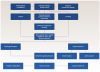
A decision tree can be beneficial in medicine and dentistry for managing patients in a logical and efficient way.1 The use of decision trees is a reliable technique that provides high classification accuracy with a simple representation of state-of-the-art knowledge in different areas of medical decision-making.1 While the present authors have previously published a surgical/orthodontic decision tree for managing impacted maxillary canines,2 this article presents a surgical/orthodontic decision tree for the management of impacted maxillary central incisors (Figure 1).
Impacted maxillary central incisors represent a complex problem with functional and esthetic consequences in younger individuals, whose personality is still being developed.3 In roughly 65% of such cases, the impaction is related to trauma the child received in the anterior portion of the mouth during primary dentition. Facial trauma can produce displacement of the permanent maxillary central incisor tooth germ. Other instances of impacted maxillary central incisors may be due to a physical blockage that interrupts normal eruption with the local presence of supernumeraries or odontomas. Additionally, tooth size-to-arch length discrepancy can produce a lack of space in the premaxilla, disallowing the eruption of the maxillary central incisors. Lastly, in very rare instances, impactions of maxillary central incisors may be related to syndromes that involve multiple impacted and supernumerary teeth.3-5
Fortunately, impaction of maxillary central incisors is a relatively uncommon situation, affecting less than 1% of the general population.6 It is three times more common in boys than girls due to the nature of their playing activity and a greater possibility of receiving anterior dentoalveolar trauma.7 Common dentoalveolar alterations with impacted maxillary central incisors include dilacerated and altered roots in form (25% shorter root in length compared to normally erupted incisors),8 a decrease in thickness and height of labial alveolar bone in the impacted incisor relative to the homologous maxillary central incisor, and presence of gingival recessions.9 The most common complications related to impacted maxillary central incisors are ankylosis, root changes in form and size, and soft-tissue esthetic discrepancies in comparison to the non-impacted homologous maxillary central incisor. Scars in the soft tissue can significantly affect smile esthetics. Long-term follow-up to assess soft-tissue maturation is part of the treatment and retention protocol in these cases.3,7-10
Diagnosis and Management Timing
Tooth eruption typically occurs when approximately three-quarters of the root length development is completed. Maxillary central incisors typically erupt at ages 6½ to 7 years, while maxillary lateral incisors erupt around a year later. Parents, caregivers, pediatric dentists, and/or pediatricians may often notice a difference in the eruption age of one maxillary central incisor compared to the other, in which case an orthodontic consultation is suggested.7 Clinical palpation, anamnesis, and imaging, including a panoramic screening x-ray or sometimes a lateral cephalometric x-ray, might lead to the diagnosis of impaction.5-7 After this, a periapical x-ray is taken; today, more sophisticated imaging such as cone-beam computed tomography (CBCT) is commonly required to assess in three dimensions the amount of tooth displacement and the presence of blocking elements, such as odontomas or supernumeraries, and their relation to other nearby structures.7
Interdisciplinary consultations with a periodontist or oral surgeon are needed when simple measures such as space opening do not provide an efficient and prompt resolution to the problem.5 Other factors to take into account are surrounding teeth (ie, the integrity and presence of well-formed maxillary lateral incisors), the available amount of attached gingiva and overall periodontal conditions, and the presence of root dilacerations and/or shorter roots with altered eruption against the cortical plates.7,9 The presence of deciduous teeth, supernumerary teeth, and/or odontomas that might require timely surgical removal to eliminate the cause of obstruction to the eruption and/or traction is also an important consideration.3-5,7 More complex and rare cases, such as those that are syndromic in nature, might require a complete medical and genetic evaluation. Several syndromes are associated with supernumerary teeth and impacted maxillary central incisors.10
An impacted maxillary permanent central incisor requires management as soon as diagnosed; this usually occurs between ages 8 and 10 years.5,7 Despite incomplete tooth formation and even without three-quarters root completion, orthodontic tooth reorientation is of utmost importance.7 In horizontally impacted or severely displaced and/or rotated maxillary central incisors, intervention should take place as soon as the condition is identified to facilitate further root formation completion in medullary bone away from the cortical plate, because when the cortical plate establishes contact with the impacted maxillary central incisor it alters the root formation process, resulting in a tooth with a deformed and shorter root.3,5,7
Orthodontic Management
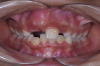
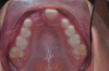
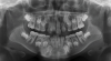
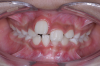
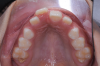
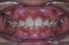
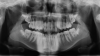
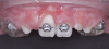
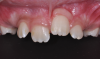
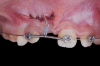
The following case illustrates the complete surgical/orthodontic treatment sequence for an impacted maxillary right central incisor. An 8½-year-old patient presented with a chief complaint of crowded teeth. The maxillary left central incisor was the only erupted incisor at the time of consultation (Figure 2 and Figure 3). Crowding was clinically diagnosed visually by the prominence of the unerupted right central incisor in the vestibule, and this was confirmed with a panoramic radiograph (Figure 4).
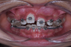
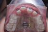
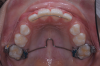
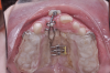
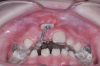
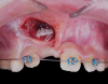
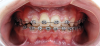
The maxillary right permanent centralincisor had a delayed eruption of 1½ years compared to its homologous incisor, and it was anteriorly displaced due to insufficient space in the premaxilla. Removal of the maxillary deciduous canines was indicated to unravel the crowding. An apically positioned flap of the impacted right central incisor was made to facilitate its eruption (Figure 5 and Figure 6). The case was worked up to plan an active phase l treatment, consisting of palatal expansion and the use of sectional fixed appliances in the upper arch. A transverse deficiency without posterior crossbite was identified, and therefore palatal expansion with a bonded expander was planned to widen the premaxilla (Figure 7 and Figure 8). A two-by-four fixed appliance system (brackets in the four maxillary anterior teeth and bands on the maxillary first molars) was used to align the maxillary teeth. The gingival margin of the retained maxillary right central incisor was slightly more apical than the left one, which erupted as expected (Figure 9 and Figure 10). Figure 11 shows a panoramic radiograph taken after completion of phase 1 treatment.
In reviewing this case more closely, the sequence usually starts with a space opening (Figure 12 through Figure 14). Palatal expansion is generally performed even without the presence of a posterior dental crossbite, as many patients with impacted maxillary central incisors are deficient in the development of the anterior portion of the maxilla.7,11 The use of sliding mechanics with open coils in conjunction with simple fixed systems is preferred to removable appliances since they deliver a light, continued force instead of an intermittent force.7 Interarch mechanics allow space to be gained for either the natural eruption or the surgical/orthodontic traction of the impacted maxillary central incisor.7 After palatal expansion, rigid structures, such as fixed transpalatal arches soldered to upper first molar bands, might first be used to anchor the active traction of the impacted tooth. Later, orthodontic fixed appliances reportedly have been used in the maxillary anterior to detail the position of the maxillary central incisor in the arch.11 Frequently, power arms are incorporated into palatal expanders.12 Fixed systems initially employ flexible nitinol wires and round stainless-steel wires. After initial leveling, rectangular stainless-steel wires have been used as part of several protocols to produce torque and place the roots of the resolved impacted central incisor in the proper position in its apical base.3,7,13 Orthodontic traction is conducted with different systems based on the preference of the clinician.3,7,10
Surgical Exposure Options for Impacted Teeth
Pavoni et al compiled several maxillary central impaction orientation methods for assessing the difficulty in surgical and orthodontic management based on panoramic x-ray (initial screening imaging).14 Their criteria for locating impacted maxillary central incisors included the impacted incisor inclination to the midline, the incisor's occlusal-gingival height relative to its homologous incisor, and its distance to the occlusal plane, as well as the amount of the impacted tooth root formation. More complex imaging, which might include a CBCT scan with a lip retractor to separate the cheeks from the buccal surfaces of the teeth, is usually required. This allows a more precise determination of bone height relative to the cementoenamel junction.7 Complete clinical and imaging information is helpful in the presence of physical blockage or severe incisor displacement if supernumerary teeth or odontomas are the cause.7-9
Open-Eruption Technique
The open-eruption technique involves repositioning the soft tissue and removing bone to expose the crown. This technique is recommended when the impacted tooth is in a high position with no attached gingiva, such as seen in Figure 15.7,15 The goal is to ensure a keratinized gingival margin for proper alignment and prevent future gingival recession and bone loss. Cases treated with apically positioned flaps benefit esthetically as the gingival tissue matures over time.7,15 Additionally, this approach provides the orthodontist a direct visualization, simplifying traction. If the orthodontic attachment becomes loose, there is no need to uncover the tooth again.15 Critics, however, claim this technique may result in slight reintrusion, irregular gingival contours, and scar tissue post-treatment.16
Closed-Eruption Technique
On the other hand, the closed-eruption technique aims to cover the tooth with a flap and bond an attachment to resemble a natural eruption process (Figure 16 and Figure 17).16 Proponents of this technique report that more than 90% of impacted maxillary central incisors are buccally oriented and view this technique as advantageous, as the closed eruption mimics normal tooth emergence.7,16
Opponents of the closed-eruption technique criticize the higher risk of ankylosis and slower speed of movement of the impacted incisor. Because the flap is closed after the uncovering, the orthodontic bonded attachment might require a second surgical exposure if it becomes loose.7,15 Also, a shorter height of the attached gingiva at the impacted tooth as compared to the normally erupted incisor may result.7 Some impacted maxillary central incisors are so high in the vestibule that a closed-eruption technique might be required first, followed by an apically positioned flap.3
Managing Complications
Complications such as root dilaceration, short roots, and ankylosis often require a multidisciplinary approach involving orthodontic, periodontal, and/or oral surgeon specialists and restorative dentists. Treatment strategies for these complications may encompass surgical interventions, orthodontic realignment, bone grafting, or the use of orthodontic mechanics to achieve a compromise outcome.7-9,17-19 Early intervention can help identify these complications, enabling their timely management and minimizing potential long-term consequences. Orthodontic and periodontal follow-ups are part of the retention protocols.7,17-19
Root dilaceration, which refers to the abnormal curvature or bending of the tooth's root, can complicate orthodontic treatment, necessitating specialized techniques to navigate the curved or bent root structure in an attempt to place the root in the apical base.7,19 Short roots can present a challenge, as they may not provide adequate support for the tooth, potentially leading to instability or premature tooth loss (Figure 18).7-9
Ankylosis occurs when the tooth becomes fused to the surrounding bone, inhibiting normal tooth movement and potentially causing functional and esthetic issues.20 Ankylosis can result in altered tooth eruption patterns and abnormal occlusal relationships, requiring additional surgical and/or orthodontic intervention to correct functional and esthetic concerns.7,20
Periodontal soft-tissue scars or deformities have been reported when comparing an impacted maxillary central incisor with its homologous non-impacted counterpart (Figure 19).9,21
Final Considerations
Maxillary central incisors are crucial for phonetics, eating, and dentofacial esthetics. Impacted maxillary central incisors can be managed effectively through a comprehensive surgical and orthodontic approach. Understanding the causes of impaction, establishing a suitable treatment plan through a comprehensive clinical and imaging diagnosis, and using sound orthodontic biomechanical principles and an adequate soft-tissue surgical technique are essential for success. The orthodontist plays a critical role in designing the different lever arms that can be integrated into appliances such as expanders or other soldered fixed appliances. Long-term periodontal follow-up of the impacted maxillary central incisor should be seen as part of continuing active treatment to secure good periodontal health and adequate esthetics. Other treatment alternatives in selective cases, such as autotransplants, decoronation, lateral incisor or canine substitution with the aid of esthetic dentistry or implant replacement, may sometimes be required.
About the Authors
Miguel Hirschhaut, DDS, PhD
Private Practice limited to Orthodontics, Caracas, Venezuela
Carol Weinstein, DDS
Private Practice limited to Orthodontics, Santiago de Chile, Chile
Carolina Alarcon, DDS
Private Practice limited to Periodontics, Santiago de Chile, Chile
Carlos Flores-Mir, DDS, DSc
Professor, Division of Orthodontics, Department of Dentistry, University of Alberta, Edmonton, Alberta, Canada; Part-time Private Practice limited to Orthodontics, Edmonton, Alberta, Canada
Queries to the author regarding this course may be submitted to authorqueries@broadcastmed.com.
References
1. Podgorelec V, Kokol P, Stiglic B, Rozman I. Decision trees: an overview and their use in medicine. J Med Syst. 2002;26(5):445-463.
2. Hirschhaut M, Leon N, Gross H, Flores-Mir C. Guidance for the clinical management of impacted maxillary canines. Compend Contin Educ Dent. 2021;42(5):220-226.
3. Chaushu S, Becker T, Becker A. Impacted central incisors: factors affecting prognosis and treatment duration. Am J Orthod Dentofac Orthop. 2015;147(3):355-362.
4. Seehra J, Mortaja K, Wazwaz F, et al. Interventions to facilitate the successful eruption of impacted maxillary incisor teeth due to the presence of a supernumerary: a systematic review and meta-analysis. Am J Orthod Dentofacial Orthop. 2023;163(5):594-608.
5. Becker A. Early treatment for impacted maxillary incisors. Am J Orthod Dentofacial Orthop. 2002;121(6):586-587.
6. Bartolo A, Camilleri A, Camilleri S. Unerupted incisors-characteristic features and associated anomalies. Eur J Orthod. 2010;32(3):297-301.
7. Weinstein C, Alarcón C, Flores-Mir C, et al. Orthodontic and periodontal considerations in maxillary central incisor impactions: case series. Am J Orthod Dentofacial Orthop Clinical Companion. 2022;2(4):372-386.
8. Lana Mattiello FD, Deon Rizzatto SM, Macedo de Menezes L, et al. Dimensional and morphologic characteristics of unilateral impacted maxillary central incisors. Am J Orthod Dentofacial Orthop. 2022;162(3):340-347.
9. Calil LR, Janson G, Silva VMD, et al. Periodontal status of maxillary central incisors after orthodontic traction: a longitudinal follow-up. J Appl Oral Sci. 2022;30:e20210492. doi: 10.1590/1678-7757-2021-0492.
10. Lubinsky M, Kantaputra PN. Syndromes with supernumerary teeth. Am J Med Genet A. 2016;170(10):2611-2616.
11. Greco M, Pacifico D, Rombolà A. A three-stage approach for managing impacted maxillary incisors in the mixed dentition. J Clin Orthod. 2022;56(6):334-342.
12. Sant'Anna EF, Marquezan M, Sant'Anna CF. Impacted incisors associated with supernumerary teeth treated with a modified Haas appliance. Am J Orthod Dentofacial Orthop. 2012;142(6):863-871.
13. Singh H, Kapoor P, Sharma P, et al. Interdisciplinary management of an impacted dilacerated maxillary central incisor. Dental Press J Orthod. 2018;23(3):37-46.
14. Pavoni C, Mucedero M, Laganá G, et al. Impacted maxillary incisors: diagnosis and predictive measurements. Ann Stomatol (Roma). 2012;3(3-4):100-105.
15. Vanarsdall RL Jr. Efficient management of unerupted teeth: a time-tested treatment modality. Sem Orthod. 2010;16(3):212-221.
16. Kokich VG, Mathews DP. Orthodontic and Surgical Management of Impacted Teeth. 1st ed. Chicago, IL: Quintessence Publishing; 2012:1-25.
17. Campbell KM, Casas MJ, Kenny DJ. Ankylosis of traumatized permanent incisors: pathogenesis and current approaches to diagnosis and management. J Can Dent Assoc. 2005;71(10):763-768.
18. Chang NY, Park JH, Kim SC, et al. Forced eruption of impacted maxillary central incisors with severely dilacerated roots. Am J Orthod Dentofacial Orthop. 2016;150(4):692-702.
19. Pavlidis D, Daratsianos N, Jager A. Treatment of an impacted dilacerated maxillary central incisor. Am J Orthod Dentofacial Orthop. 2011;139(3):378-387.
20. Kvint S, Lindsten R, Magnusson A, et al. Autotransplantation of teeth in 215 Patients. A follow-up study. Angle Orthod. 2010;80(3):446-451.
21. Chaushu S, Dykstein N, Ben-Bassat Y, Becker A. Periodontal status of impacted maxillary incisors uncovered by 2 different surgical techniques. J Oral Maxillofac Surg. 2009;67(1):120-124.